Copyright
©2014 Baishideng Publishing Group Co.
World J Gastrointest Oncol. Jan 15, 2014; 6(1): 11-21
Published online Jan 15, 2014. doi: 10.4251/wjgo.v6.i1.11
Published online Jan 15, 2014. doi: 10.4251/wjgo.v6.i1.11
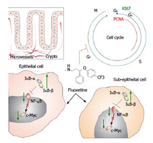
Figure 3 Schematic illustration shows fluoxetine antiproliferative activities in colon tissue.
Boxed figure shows the clear division between epithelial and subepithelial colonic areas. Considering that crypts compose the colonic epithelia, it is known that microvessels surround these gland structures. Fluoxetine (chemical structure represented at the center) blocks cell-cycle (blue line and letters) in colonic tissue. We have observed that fluoxetine treatment reduced two proliferative markers, named proliferating cell nuclear antigen (PCNA, red line) and KI67 (green line). These effects of fluoxetine treatment might be related to its enhancement on IκB-α and IκB-β proteins. This could arrest nuclear factor kappa-B (NF-κB) protein in the cytoplasm, reducing its transcriptional activity which, due to its activation over c-Myc transcription factor, would decrease this protein activation and proliferation. We believe that a similar mechanism could take a place in epithelial and subepithelial cells.
- Citation: Stopper H, Garcia SB, Waaga-Gasser AM, Kannen V. Antidepressant fluoxetine and its potential against colon tumors. World J Gastrointest Oncol 2014; 6(1): 11-21
- URL: https://www.wjgnet.com/1948-5204/full/v6/i1/11.htm
- DOI: https://dx.doi.org/10.4251/wjgo.v6.i1.11


