Copyright
©2013 Baishideng Publishing Group Co.
World J Gastrointest Oncol. Jul 15, 2013; 5(7): 115-126
Published online Jul 15, 2013. doi: 10.4251/wjgo.v5.i7.115
Published online Jul 15, 2013. doi: 10.4251/wjgo.v5.i7.115
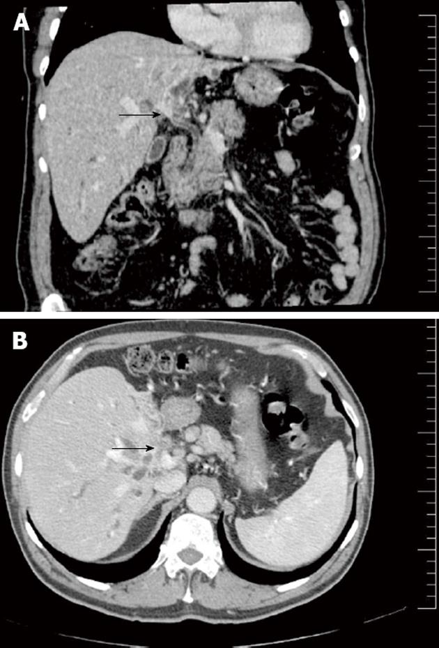
Figure 10 Liver infiltration of cholangiocarcinoma.
A: Coronal reconstructed multidetector computed tomography shows a periductal mass at the hepatic confluence consistent with cholangiocarcinoma (arrow); B: Axial computed tomography of the same patient shows a hypoenhancing mass involving the liver parenchyma and hilar vessels (arrow) consistent with tumoral infiltration by cholangiocarcinoma.
- Citation: Valls C, Ruiz S, Martinez L, Leiva D. Radiological diagnosis and staging of hilar cholangiocarcinoma. World J Gastrointest Oncol 2013; 5(7): 115-126
- URL: https://www.wjgnet.com/1948-5204/full/v5/i7/115.htm
- DOI: https://dx.doi.org/10.4251/wjgo.v5.i7.115


