Copyright
©2013 Baishideng Publishing Group Co.
World J Gastrointest Oncol. Jul 15, 2013; 5(7): 115-126
Published online Jul 15, 2013. doi: 10.4251/wjgo.v5.i7.115
Published online Jul 15, 2013. doi: 10.4251/wjgo.v5.i7.115
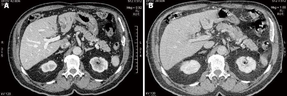
Figure 3 Periductal cholangiocarcinoma multidetector computed tomography.
A: Multidetector computed tomography (MDCT) in the portal phase at the level of the hepatic hilus shows an irregular periductal thickening completely obstructing the common hepatic duct consistent with periductal cholangiocarcinoma (arrow). The lesion is hypoenhancing in the portal phase; B: Delayed phase MDCT at the same level shows marked hyper-enhancement of the tumoral lesion (arrow).
- Citation: Valls C, Ruiz S, Martinez L, Leiva D. Radiological diagnosis and staging of hilar cholangiocarcinoma. World J Gastrointest Oncol 2013; 5(7): 115-126
- URL: https://www.wjgnet.com/1948-5204/full/v5/i7/115.htm
- DOI: https://dx.doi.org/10.4251/wjgo.v5.i7.115


