Copyright
©2013 Baishideng Publishing Group Co.
World J Gastrointest Oncol. Jul 15, 2013; 5(7): 115-126
Published online Jul 15, 2013. doi: 10.4251/wjgo.v5.i7.115
Published online Jul 15, 2013. doi: 10.4251/wjgo.v5.i7.115
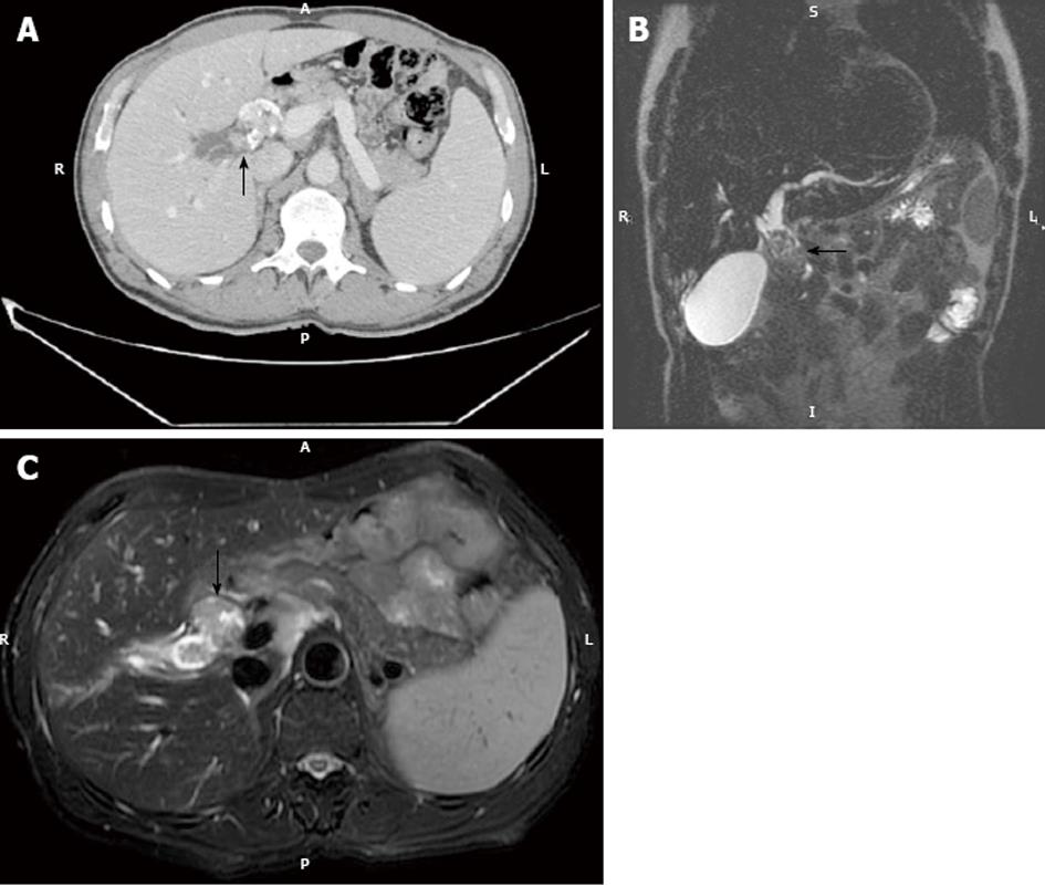
Figure 2 Intraductal growing cholangiocarcinoma.
A: Axial computed tomography in the portal phase in a 52-year-old male shows a heterogeneously enhancing endoluminal lesion with dysmorphic calcifications (arrow). Note biliary dilatation upstream; B: Coronal thin-slab (echo spacing 4.2 ms, effective echo time 183 ms, image matrix 272 x 512, FOV 385 mm) T2-W sequence shows marked dilatation of the common hepatic duct and a hypointense endoluminal mass (arrow); C: Axial thin-slab (echo spacing 4.2 ms, effective echo time 183 ms, image matrix 272 x 512, FOV 385 mm) T2-W sequence shows a hypointense filling defect consistent with polypoid cholangiocarcinoma in the common hepatic duct (arrow).
- Citation: Valls C, Ruiz S, Martinez L, Leiva D. Radiological diagnosis and staging of hilar cholangiocarcinoma. World J Gastrointest Oncol 2013; 5(7): 115-126
- URL: https://www.wjgnet.com/1948-5204/full/v5/i7/115.htm
- DOI: https://dx.doi.org/10.4251/wjgo.v5.i7.115


