Copyright
©2013 Baishideng Publishing Group Co.
World J Gastrointest Oncol. Dec 15, 2013; 5(12): 222-229
Published online Dec 15, 2013. doi: 10.4251/wjgo.v5.i12.222
Published online Dec 15, 2013. doi: 10.4251/wjgo.v5.i12.222
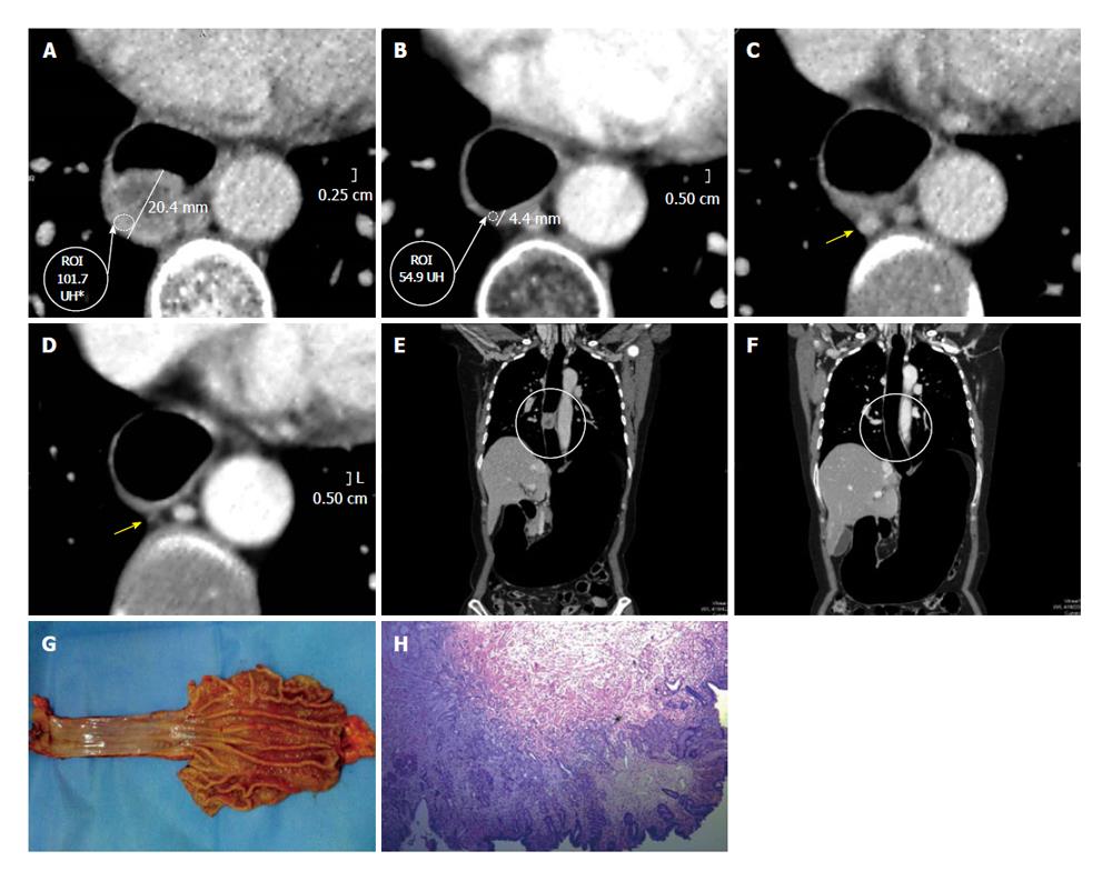
Figure 8 Epidermoid carcinoma of the thoracic esophagus [CME #5].
Dworak grade 4, absence of tumor cells. A: Axial pre-neoadjuvant therapy image. Both wall thickness and density are measured; B: Axial post- neoadjuvant therapy image reveals a clear decrease of the wall thickening and density; C: Axial pre- neoadjuvant therapy image, the arrow is pointing to adenopathy; D: Axial post-neoadjuvancy image reveals disappearance of adenopathy; E: Coronal multiplanar reconstructions (MPR) pre-neoadjuvant therapy reconstruction. The circle shows the long axis of the tumor; F: Coronal MPR reconstruction post- neoadjuvant therapy. The neoplasm is not longer detected; G: Surgical specimen of total esophagectomy and upper polar gastrectomy. Open piece, is recognized at the level of the gastro-esophageal junction an area of white-depressed with elevated edges; H: Squamous epithelium with acanthosis, conserved cell polarity. Fibrohialinosis in lamina propria and submucosa with lymphocytic infiltrate. Absence of atypical cells. At the level of the gastroesophageal junction shows a sector with intestinal metaplasia in the stomach side, negative for dysplasia.
- Citation: Ulla M, Gentile E, Yeyati EL, Diez ML, Cavadas D, Garcia-Monaco RD, Ros PR. Pneumo-CT assessing response to neoadjuvant therapy in esophageal cancer: Imaging-pathological correlation. World J Gastrointest Oncol 2013; 5(12): 222-229
- URL: https://www.wjgnet.com/1948-5204/full/v5/i12/222.htm
- DOI: https://dx.doi.org/10.4251/wjgo.v5.i12.222


