Copyright
©2013 Baishideng Publishing Group Co.
World J Gastrointest Oncol. Dec 15, 2013; 5(12): 222-229
Published online Dec 15, 2013. doi: 10.4251/wjgo.v5.i12.222
Published online Dec 15, 2013. doi: 10.4251/wjgo.v5.i12.222
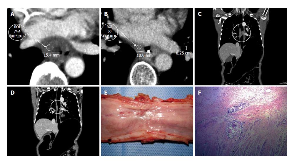
Figure 7 Epidermoid carcinoma.
Dworak grade 3, scarce neoplastic cells. A: Axial pre-neoadjuvant therapy image. Both wall thickness and density are measured; B: Axial post- neoadjuvant therapy image reveals a clear decrease of the wall thickening and density; C: Coronal multiplanar reconstructions (MPR) pre-neoadjuvant therapy reconstruction. The circle shows the long axis of the tumor; D: Coronal MPR post-neoadjuvant therapy reconstruction with decrease in tumor size; E: Surgical specimen of total esophagectomy. Open piece, shows thickening at the middle third of the esophagus with an ulcerated area; F: The sections shows at submucosal layer a nodular accumulation of atypical epithelial cells with vesicular nuclei and scant cytoplasm that are arranged in small tubular structures. Also the submucosa presents fibrosis, chronic inflammation and congestion. Mucosal layer shows conserved squamous epithelium and focal fibrosis regression suggesting changes.
- Citation: Ulla M, Gentile E, Yeyati EL, Diez ML, Cavadas D, Garcia-Monaco RD, Ros PR. Pneumo-CT assessing response to neoadjuvant therapy in esophageal cancer: Imaging-pathological correlation. World J Gastrointest Oncol 2013; 5(12): 222-229
- URL: https://www.wjgnet.com/1948-5204/full/v5/i12/222.htm
- DOI: https://dx.doi.org/10.4251/wjgo.v5.i12.222


