Copyright
©2012 Baishideng Publishing Group Co.
World J Gastrointest Oncol. Sep 15, 2012; 4(9): 202-206
Published online Sep 15, 2012. doi: 10.4251/wjgo.v4.i9.202
Published online Sep 15, 2012. doi: 10.4251/wjgo.v4.i9.202
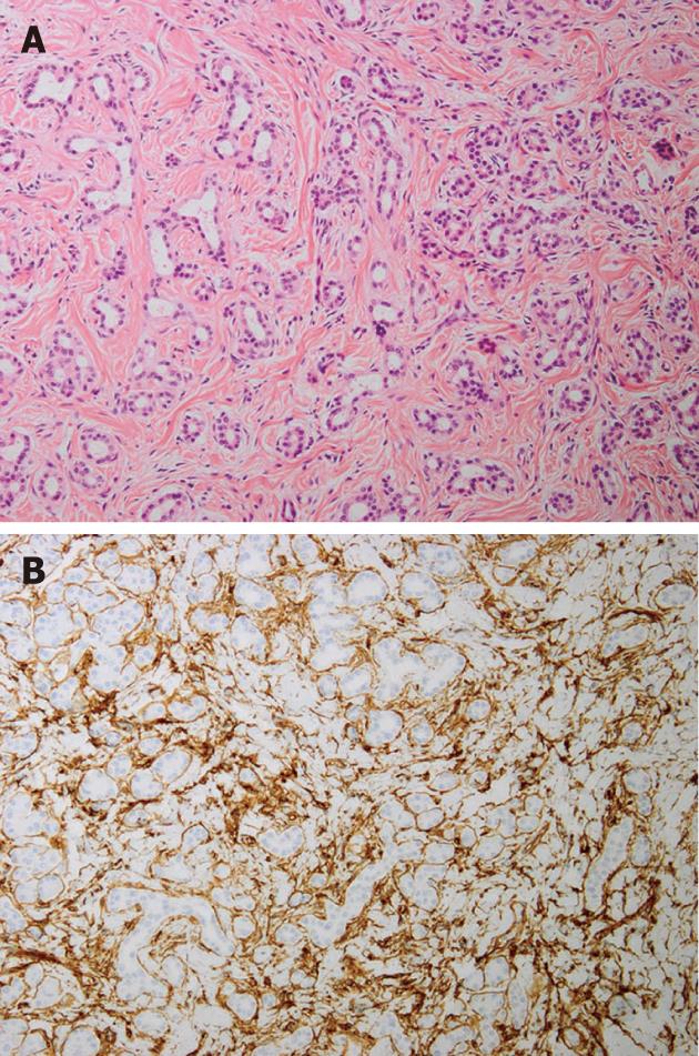
Figure 3 Microscopic images of the tumors.
A: The lesion is composed of non-neoplastic acinar and ductal cells embedded in hypocellular fibrous stroma (HE stain, 100 x); B: Immunohistochemically, stromal spindle cells are positive for CD34 (100 x).
- Citation: Kawakami F, Shimizu M, Yamaguchi H, Hara S, Matsumoto I, Ku Y, Itoh T. Multiple solid pancreatic hamartomas: A case report and review of the literature. World J Gastrointest Oncol 2012; 4(9): 202-206
- URL: https://www.wjgnet.com/1948-5204/full/v4/i9/202.htm
- DOI: https://dx.doi.org/10.4251/wjgo.v4.i9.202


