Copyright
©2012 Baishideng.
World J Gastrointest Oncol. Apr 15, 2012; 4(4): 89-93
Published online Apr 15, 2012. doi: 10.4251/wjgo.v4.i4.89
Published online Apr 15, 2012. doi: 10.4251/wjgo.v4.i4.89
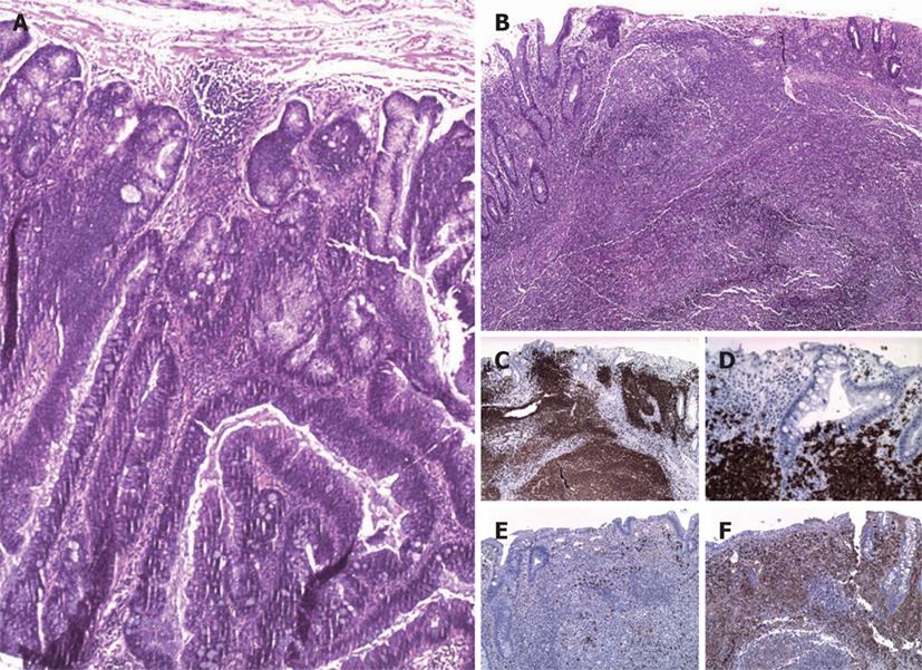
Figure 1 Histological examination of Case 1.
Tubulovillous adenoma with superficial foci of high grade intraepithelial neoplasia (A: HE, × 4); coexisting colonic lymphoma of MALT type [B: HE, × 4; C: CD20, × 4; D: CD20, × 20 (lymphoepithelial lesions); E: κ light chain, × 4; F: λ light chain, × 4].
- Citation: Argyropoulos T, Foukas P, Kefala M, Xylardistos P, Papageorgiou S, Machairas N, Boltetsou E, Machairas A, Panayiotides IG. Simultaneous occurrence of colonic adenocarcinoma and MALT lymphoma: A series of three cases. World J Gastrointest Oncol 2012; 4(4): 89-93
- URL: https://www.wjgnet.com/1948-5204/full/v4/i4/89.htm
- DOI: https://dx.doi.org/10.4251/wjgo.v4.i4.89


