Copyright
©2012 Baishideng Publishing Group Co.
World J Gastrointest Oncol. Dec 15, 2012; 4(12): 238-249
Published online Dec 15, 2012. doi: 10.4251/wjgo.v4.i12.238
Published online Dec 15, 2012. doi: 10.4251/wjgo.v4.i12.238
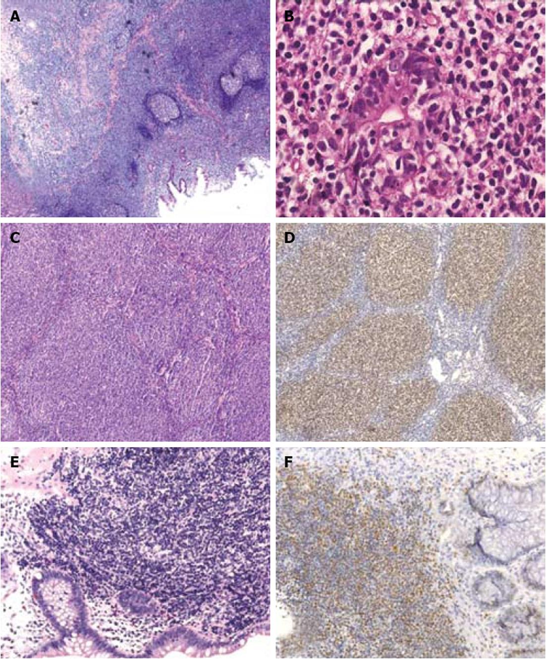
Figure 2 Histology of gastrointestinal mucosa-associated lymphoid tissue, follicular and mantle-cell lymphomas.
A: Hematoxylin eosin (HE) staining of a gastric mucosa-associated lymphoid tissue lymphoma demonstrates presence of reactive follicles surrouned by a neoplastic lymphoid infiltrate (magnification 50 ×); B: Destruction of a gastric gland by the neoplastic B-cellss: lymphoepithelial lesion (magnification 400 ×); C: HE staining of a duodenal follicular lymphoma highlights the presence of aberrant follicular growth pattern (magnification 100 ×); D: Aberrant B-cell-lymphoma-2 expression by a duodenal follicular lymphoma (magnification 100 ×); E: HE staining of a mantle-cell lymphoma in the colon (magnification 200 ×); F: Aberrant cyclin D1 expression by an intestinal mantle-cell lymphoma (magnification 100 ×).
- Citation: Sagaert X, Tousseyn T, Yantiss RK. Gastrointestinal B-cell lymphomas: From understanding B-cell physiology to classification and molecular pathology. World J Gastrointest Oncol 2012; 4(12): 238-249
- URL: https://www.wjgnet.com/1948-5204/full/v4/i12/238.htm
- DOI: https://dx.doi.org/10.4251/wjgo.v4.i12.238


