Copyright
©2013 Baishideng Publishing Group Co.
World J Gastrointest Oncol. Nov 15, 2012; 4(11): 216-222
Published online Nov 15, 2012. doi: 10.4251/wjgo.v4.i11.216
Published online Nov 15, 2012. doi: 10.4251/wjgo.v4.i11.216
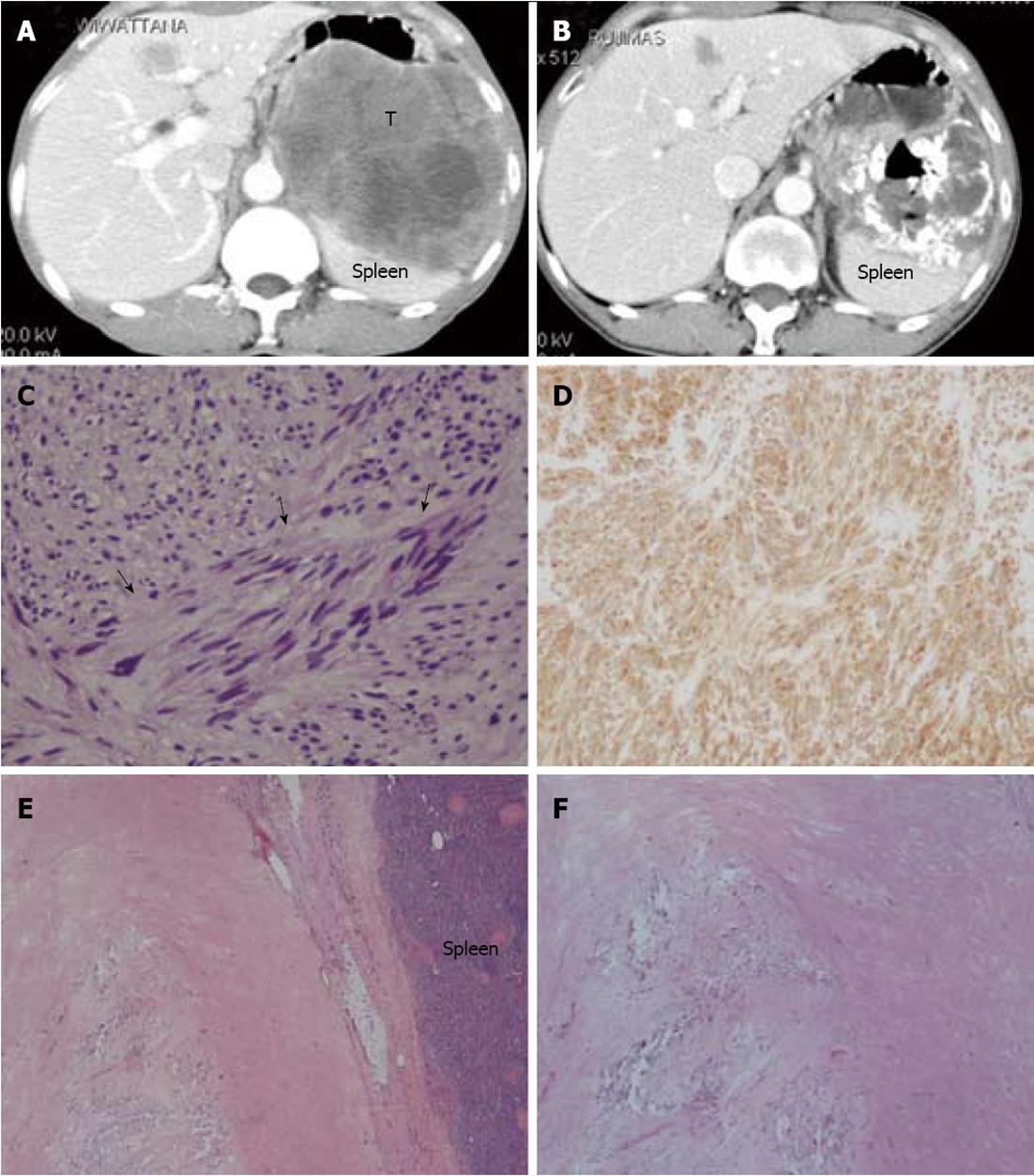
Figure 2 Abdominal computerized tomography and histopathological pictures of a 68-year-old male patient who presented with abdominal mass.
A: Computerized tomography (CT) shows a large enhancing solid mass (T), measuring 11.2 cm × 11.9 cm × 10.7 cm, occupying the left upper quadrant between the stomach and the spleen. A liver nodule is also visible in segment IV; B: Forty months following the beginning of imatinib therapy, a follow-up CT showed partial tumor response; C, D: Image-guided tissue biopsy revealed a spindle cell tumor (arrows) that marked CD117. The mitotic cell count was 2 cells/50 high power fields; E, F: Following an en bloc resection including a total removal of the stomach together with the spleen and a wedge resection of hepatic metastasis, the pathological tissue showed only a stromal hyalinization and dystrophic calcification with a scanty number of differentiated spindle cells that marked S-100, but not CD117.
- Citation: Pornsuksiri K, Chewatanakornkul S, Kanngurn S, Maneechay W, Chaiyapan W, Sangkhathat S. Clinical outcomes of gastrointestinal stromal tumor in southern Thailand. World J Gastrointest Oncol 2012; 4(11): 216-222
- URL: https://www.wjgnet.com/1948-5204/full/v4/i11/216.htm
- DOI: https://dx.doi.org/10.4251/wjgo.v4.i11.216


