Copyright
©2010 Baishideng Publishing Group Co.
World J Gastrointest Oncol. Sep 15, 2010; 2(9): 360-363
Published online Sep 15, 2010. doi: 10.4251/wjgo.v2.i9.360
Published online Sep 15, 2010. doi: 10.4251/wjgo.v2.i9.360
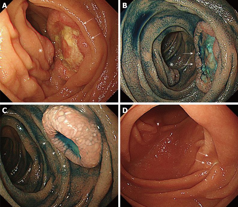
Figure 2 Esophagogastroduodenoscopy images of duodenal tumors.
A: There are two tumors at the second portion of the duodenum. Largest one (arrow) shows broadly elevated ulceration with central protrusion. Another tumor is located at the opposite side of the largest one (asterisk); B: After indigo carmine dye, ulcer base reveals a non-structural mucosal pattern with white coats. The edge of the tumor appears to be covered by normal-looking mucosa. Another tumor is located at the opposite side of the largest one (asterisk); C: Smallest one (arrowhead) is located at the most anal side. All three tumors have common features of a so-called submucosal tumor; D: After three courses of gemcitabine, three tumors at the second portion of the duodenum disappeared and are only discernible as scars in esophagogastroduodenoscopy (EGD). The scar that is derived from largest one (arrows) and another derived from the tumor located at the opposite side of the largest one (arrowheads) are discernible. The scar that is derived from the smallest is hard to see in this view of EGD.
- Citation: Miura T, Shimaoka Y, Nakamura J, Yamada S, Miura T, Yanagi M, Sato K, Usuda H, Emura I, Takahashi T. TTF-1 is useful for primary site determination in duodenal metastasis. World J Gastrointest Oncol 2010; 2(9): 360-363
- URL: https://www.wjgnet.com/1948-5204/full/v2/i9/360.htm
- DOI: https://dx.doi.org/10.4251/wjgo.v2.i9.360


