Copyright
©2010 Baishideng.
World J Gastrointest Oncol. May 15, 2010; 2(5): 222-228
Published online May 15, 2010. doi: 10.4251/wjgo.v2.i5.222
Published online May 15, 2010. doi: 10.4251/wjgo.v2.i5.222
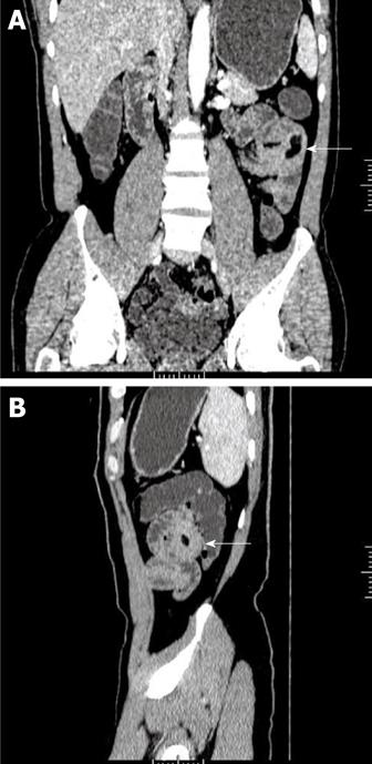
Figure 3 Lipoma of the jejunum in a 56-year-old man who presented with a 3-year history of hematochezia.
A: Image (Coronal MPR) of contrast-enhanced CT scan shows a homogeneous fat-tissue density mass in jejunal smooth muscle (white arrow); B: Image (Sagittal MPR) shows obviously thickened intestinal wall (white arrow). MPR: Multiplanar reconstruction.
- Citation: Miao F, Wang ML, Tang YH. New progress in CT and MRI examination and diagnosis of small intestinal tumors. World J Gastrointest Oncol 2010; 2(5): 222-228
- URL: https://www.wjgnet.com/1948-5204/full/v2/i5/222.htm
- DOI: https://dx.doi.org/10.4251/wjgo.v2.i5.222


