Copyright
©The Author(s) 2025.
World J Gastrointest Oncol. Apr 15, 2025; 17(4): 104919
Published online Apr 15, 2025. doi: 10.4251/wjgo.v17.i4.104919
Published online Apr 15, 2025. doi: 10.4251/wjgo.v17.i4.104919
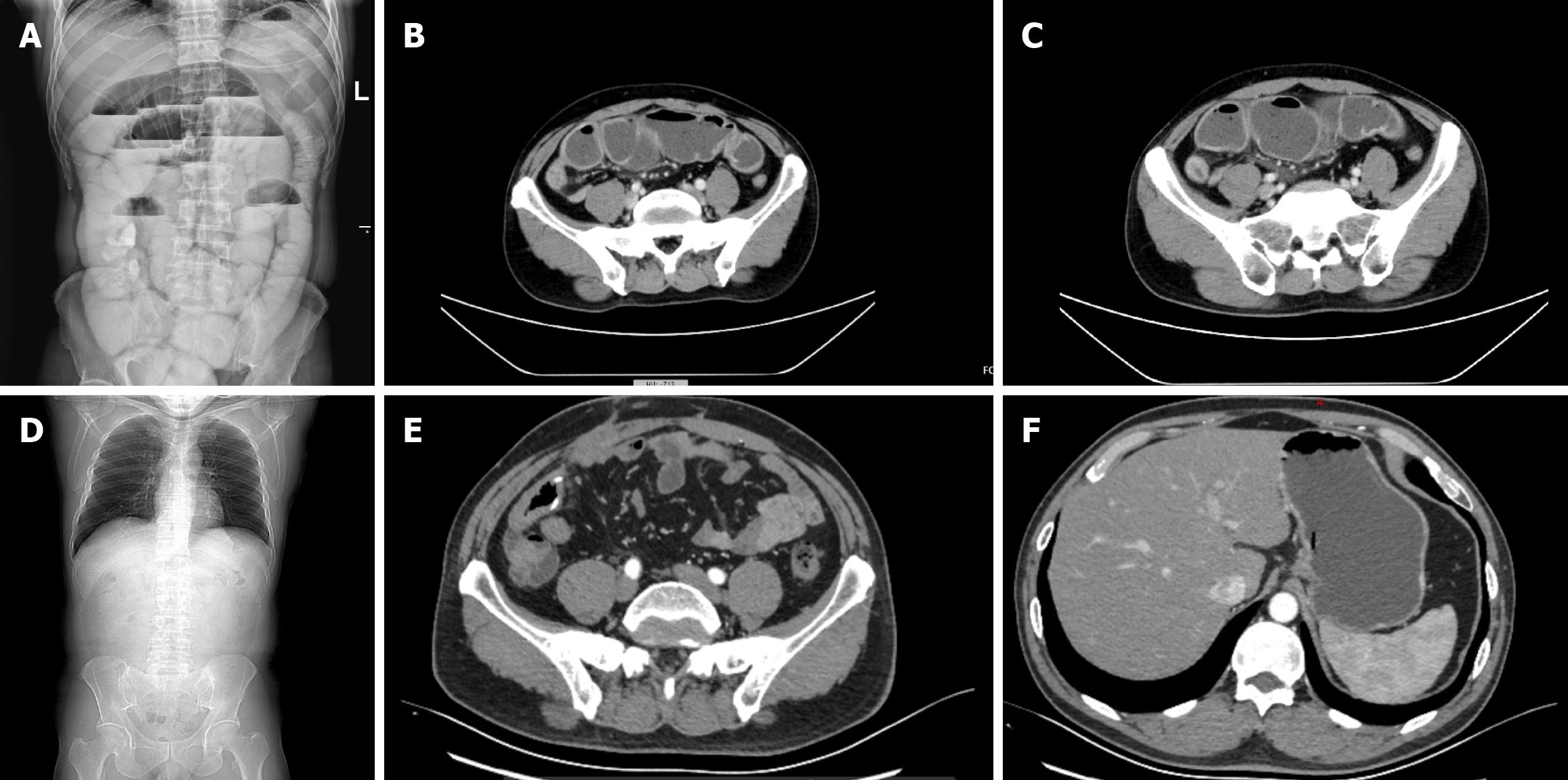
Figure 1 Digital radiology and computed tomography images of the abdomen.
A: Gas and fluid accumulation in the abdomen; B: Computed tomography (CT) scan showing intestinal obstruction; C: Uneven thickening of the ileum wall; D-F: Abdominal CT images after surgery. Two months after surgery, no recurrence or metastasis was observed under CT.
- Citation: Zhang XY, Li C, Lin J, Zhou Y, Shi RZ, Wang ZY, Jiang HB, Wang YY. Intestinal obstruction caused by early stage primary ileum adenocarcinoma: A case report and review of literature. World J Gastrointest Oncol 2025; 17(4): 104919
- URL: https://www.wjgnet.com/1948-5204/full/v17/i4/104919.htm
- DOI: https://dx.doi.org/10.4251/wjgo.v17.i4.104919


