Copyright
©The Author(s) 2025.
World J Gastrointest Oncol. Apr 15, 2025; 17(4): 103591
Published online Apr 15, 2025. doi: 10.4251/wjgo.v17.i4.103591
Published online Apr 15, 2025. doi: 10.4251/wjgo.v17.i4.103591
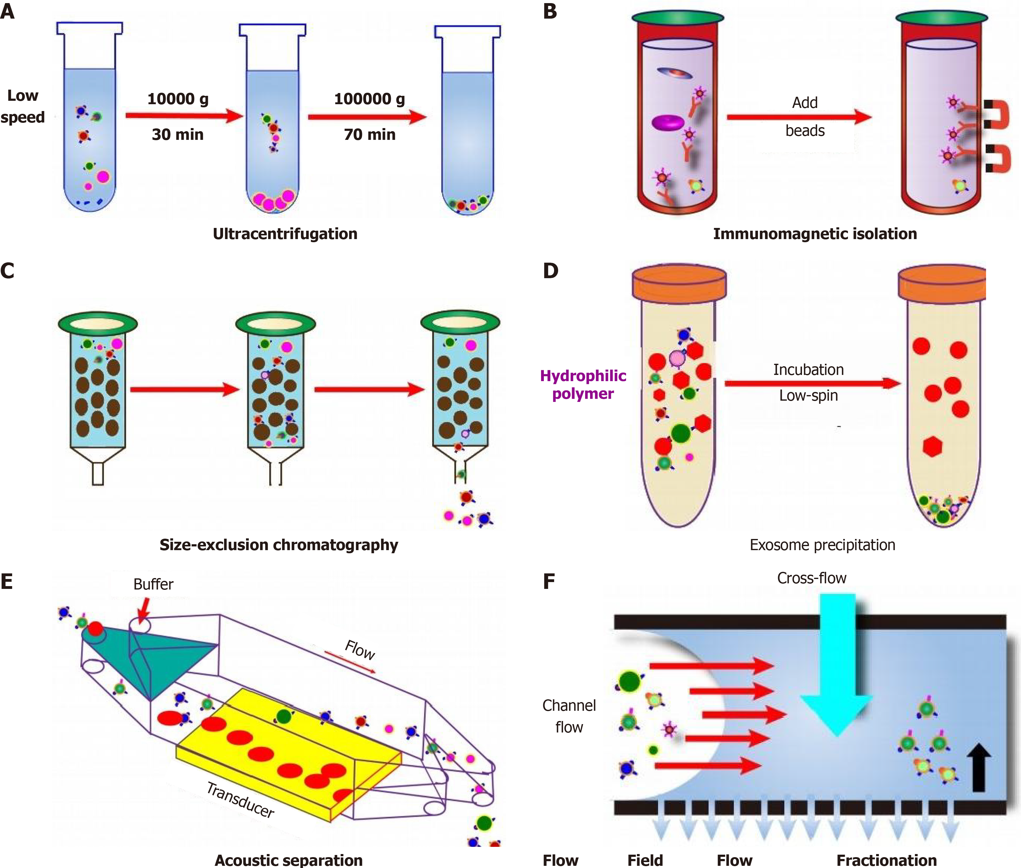
Figure 2 Schematic diagram of extracellular vesicles isolation methods and principles.
A: Ultracentrifugation is used to separate extracellular vesicles based on the precipitation subjected to their size/density; B: Using immunoaffinity magnetic beads are added to the exosome mixture and the beads with captured extracellular vesicles are separated by magnets; C: Particles of different sizes are compressed through the porous matrix and pass through the column at different speeds; D: Addition of a precipitation reagent induces that exosomes can be separated by centrifugation at a lower speed; E and F: Acoustic-based microfluidic device (E) and flow field flow fractionation (F) take advantage of the unique biophysical properties of exosomes that is capable of high-throughput separation of exosome based on field microfluidic devices use external forces (e.g., surface acoustic waves) or physical barriers to completely separate exosomes along the flow direction. Large particles are deflected to one side, while smaller exosomes are deflected to the other side.
- Citation: Zhang Y, Yue NN, Chen LY, Tian CM, Yao J, Wang LS, Liang YJ, Wei DR, Ma HL, Li DF. Exosomal biomarkers: A novel frontier in the diagnosis of gastrointestinal cancers. World J Gastrointest Oncol 2025; 17(4): 103591
- URL: https://www.wjgnet.com/1948-5204/full/v17/i4/103591.htm
- DOI: https://dx.doi.org/10.4251/wjgo.v17.i4.103591


