Copyright
©The Author(s) 2025.
World J Gastrointest Oncol. Apr 15, 2025; 17(4): 103311
Published online Apr 15, 2025. doi: 10.4251/wjgo.v17.i4.103311
Published online Apr 15, 2025. doi: 10.4251/wjgo.v17.i4.103311
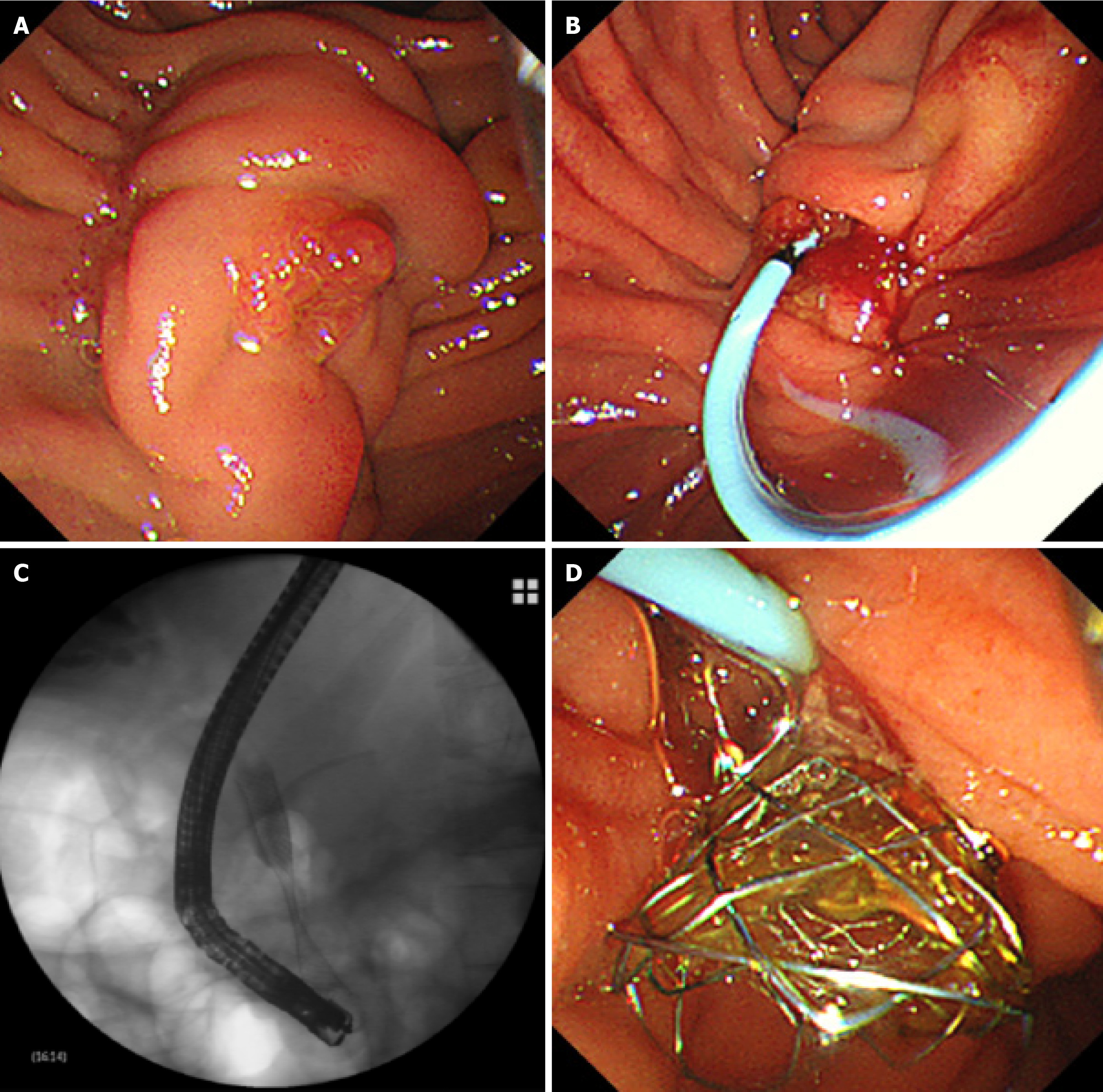
Figure 1 Endoscopic procedure.
A: The duodenal papilla can be seen under duodenoscope; B: After successful pancreatic duct cannulation, a pancreatic stent duct was placed through the guidewire; C: Radiological image of the self-expandable metallic biliary stent after deployment; D: The pancreatic stent duct and self-expandable metallic biliary stent were successfully placed under duodenoscope.
- Citation: Sun MH, Shen HZ, Jin HB, Yang JF, Zhang XF. Efficacy and safety of early pancreatic duct stenting for unresectable pancreatic cancer: A randomized controlled trial. World J Gastrointest Oncol 2025; 17(4): 103311
- URL: https://www.wjgnet.com/1948-5204/full/v17/i4/103311.htm
- DOI: https://dx.doi.org/10.4251/wjgo.v17.i4.103311


