Copyright
©The Author(s) 2025.
World J Gastrointest Oncol. Apr 15, 2025; 17(4): 101123
Published online Apr 15, 2025. doi: 10.4251/wjgo.v17.i4.101123
Published online Apr 15, 2025. doi: 10.4251/wjgo.v17.i4.101123
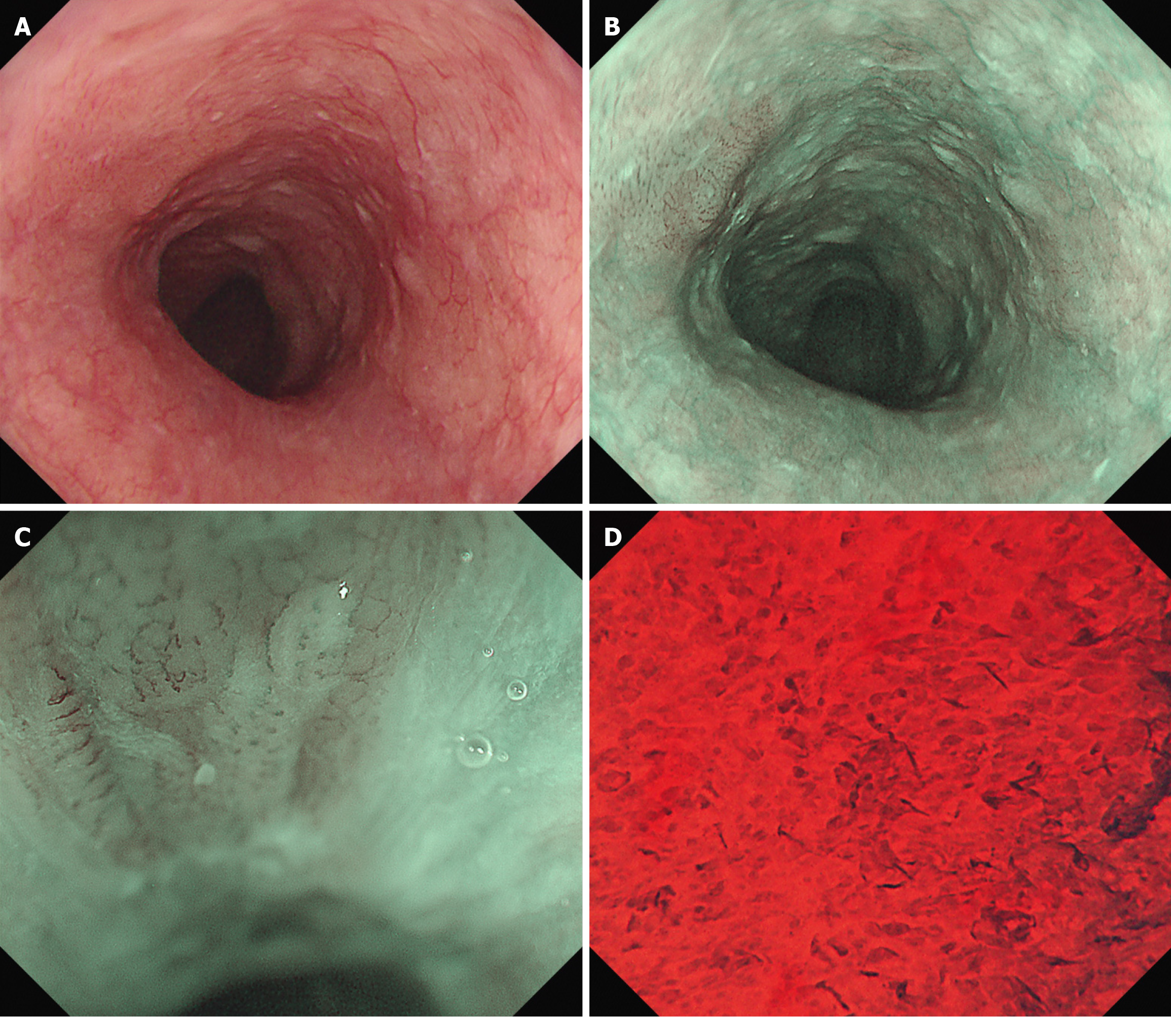
Figure 4 Endoscopic examination (after undergoing chemoradiotherapy to the neck area).
A: White light endoscopy; B: Narrow band imaging (NBI) endoscopy; C: NBI endoscopy showed type B1 vessels and small avascular area; D: Endocytoscopy showed complete loss of cellular structure with a significant increase in cellular density (EC classification: EC3). Esophagogastroduodenoscopy revealed the previously prominent elevated area had flattened and transformed into a 0-Ⅱb lesion.
- Citation: Tachibana S, Moriichi K, Takahashi K, Sato M, Kobayashi Y, Sugiyama Y, Sasaki T, Sakatani A, Ando K, Ueno N, Kashima S, Tanabe H, Fujiya M. Curative endoscopic submucosal dissection for esophageal squamous cell carcinoma after chemoradiotherapy for pharyngeal cancer: A case report. World J Gastrointest Oncol 2025; 17(4): 101123
- URL: https://www.wjgnet.com/1948-5204/full/v17/i4/101123.htm
- DOI: https://dx.doi.org/10.4251/wjgo.v17.i4.101123


