Copyright
©The Author(s) 2025.
World J Gastrointest Oncol. Apr 15, 2025; 17(4): 101123
Published online Apr 15, 2025. doi: 10.4251/wjgo.v17.i4.101123
Published online Apr 15, 2025. doi: 10.4251/wjgo.v17.i4.101123
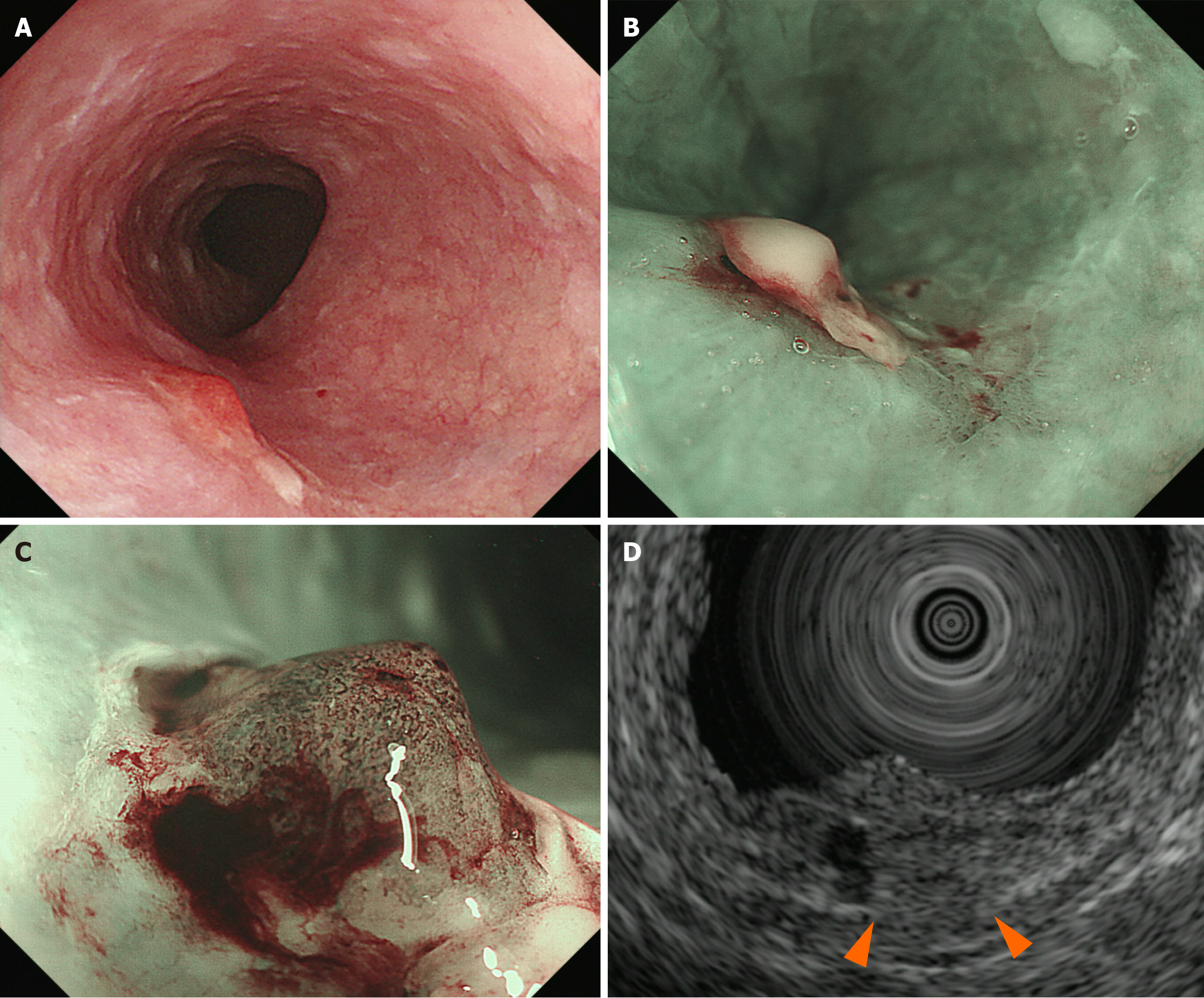
Figure 2 Endoscopic examination.
A: White light endoscopy; B: Narrow band imaging (NBI) endoscopy; C: NBI endoscopy showed type B2 vessels in prominent elevated area; D: Endoscopic ultrasonography revealed submucosal layer thinning (the fifth layer) (UM-3R 20MHz). Esophageal cancer was observed at the upper thoracic esophagus (23 cm from the incisors).
- Citation: Tachibana S, Moriichi K, Takahashi K, Sato M, Kobayashi Y, Sugiyama Y, Sasaki T, Sakatani A, Ando K, Ueno N, Kashima S, Tanabe H, Fujiya M. Curative endoscopic submucosal dissection for esophageal squamous cell carcinoma after chemoradiotherapy for pharyngeal cancer: A case report. World J Gastrointest Oncol 2025; 17(4): 101123
- URL: https://www.wjgnet.com/1948-5204/full/v17/i4/101123.htm
- DOI: https://dx.doi.org/10.4251/wjgo.v17.i4.101123


