Copyright
©The Author(s) 2025.
World J Gastrointest Oncol. Apr 15, 2025; 17(4): 100526
Published online Apr 15, 2025. doi: 10.4251/wjgo.v17.i4.100526
Published online Apr 15, 2025. doi: 10.4251/wjgo.v17.i4.100526
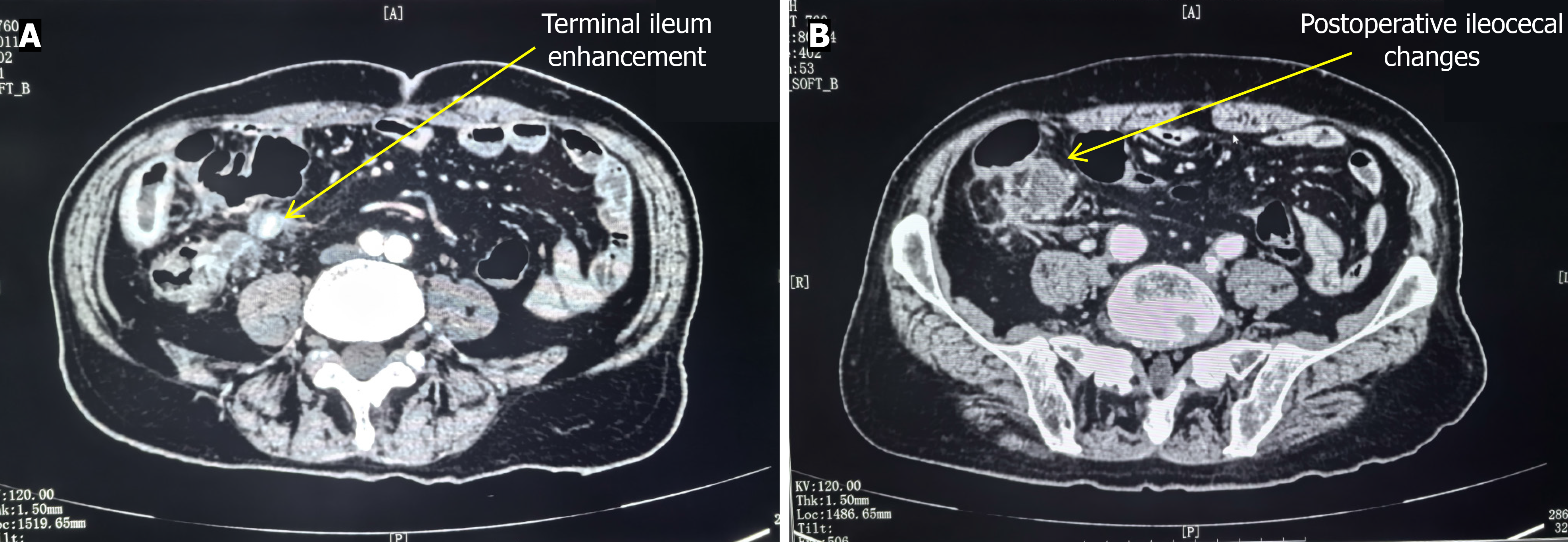
Figure 3 Imaging data of contrast-enhanced computed tomography.
A: Contrast-enhanced computed tomography scan of the abdomen showed nodular soft tissue density shadows in the operating area of the ileocecal junction, and the small intestine in the right lower abdomen was thickened and showed enhancement findings; B: After appendectomy, imaging results showed slightly dilated distal ileum, long sigmoid colon, accumulation to the right lower abdomen, and dense adhesions to the ileocecal junction and terminal ileum.
- Citation: Tang XW, Zhou Y. Signet ring cell carcinoma of the appendix and terminal ileum: A case report. World J Gastrointest Oncol 2025; 17(4): 100526
- URL: https://www.wjgnet.com/1948-5204/full/v17/i4/100526.htm
- DOI: https://dx.doi.org/10.4251/wjgo.v17.i4.100526


