Copyright
©The Author(s) 2025.
World J Gastrointest Oncol. Apr 15, 2025; 17(4): 100497
Published online Apr 15, 2025. doi: 10.4251/wjgo.v17.i4.100497
Published online Apr 15, 2025. doi: 10.4251/wjgo.v17.i4.100497
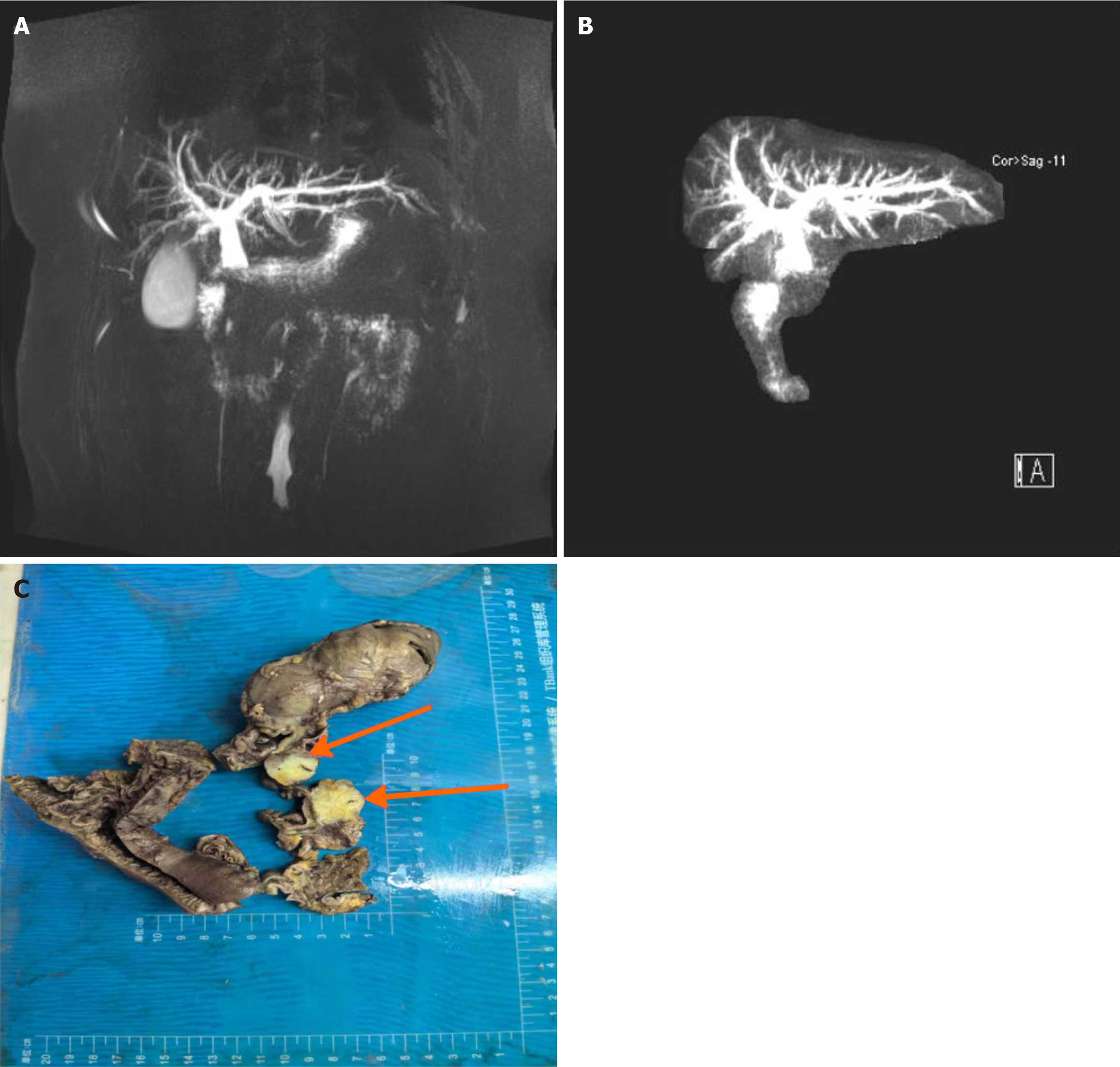
Figure 2 Imaging and pathological results of the affected area.
A and B: Magnetic resonance cholangiopancreatography images of the mass in the pancreatic head region; C: Macroscopic specimen after pathological processing. The imaging examinations were performed using a 3.0 T magnetic resonance imaging scanner for magnetic resonance cholangiopancreatography scanning, and the pathological specimens underwent standard processing procedures, including fixation, dehydration, transparency, embedding, sectioning, and hematoxylin and eosin staining. The imaging data were analyzed by two radiology experts, and the pathological specimens were evaluated macroscopically and microscopically by pathology specialists.
- Citation: Yi AQ, Xie GH. Pancreatic neuroendocrine neoplasms coexisting with biliary intraductal papillary mucinous neoplasm: A case report and review of literature. World J Gastrointest Oncol 2025; 17(4): 100497
- URL: https://www.wjgnet.com/1948-5204/full/v17/i4/100497.htm
- DOI: https://dx.doi.org/10.4251/wjgo.v17.i4.100497


