Copyright
©The Author(s) 2025.
World J Gastrointest Oncol. Mar 15, 2025; 17(3): 102083
Published online Mar 15, 2025. doi: 10.4251/wjgo.v17.i3.102083
Published online Mar 15, 2025. doi: 10.4251/wjgo.v17.i3.102083
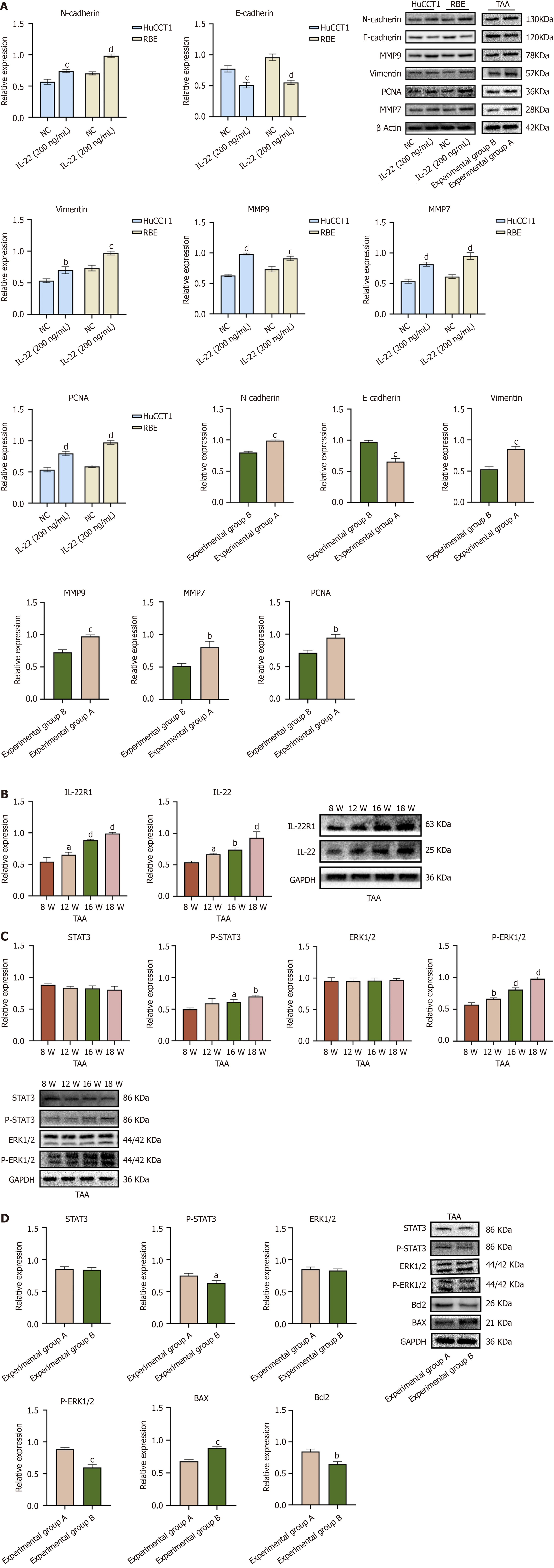
Figure 5 Protein expression levels in cholangiocarcinoma cells and tissues.
A: Western blot expression levels and quantitative analysis of related oncogenic proteins in HuCCT1 and RBE cells treated with IL-22 compared to the untreated group. Protein expression levels of related oncogenic proteins in thioacetamide (TAA) experimental groups A and B, along with the quantitative analysis results between the two groups; B: Western blot expression levels of IL-22 and IL-22R1 in liver tissues of rats at different time points following oral administration of TAA, and the Western blot quantitative analysis results comparing protein expression levels at each time point with those at the 8th week of TAA administration; C: Western blot expression levels of p-ERK1/2 and p-STAT3 in liver tissue proteins of rats administered TAA for different weeks, and the Western blot quantitative analysis results comparing protein expression levels at different weeks with those at the 8th week of TAA administration; D: Expression levels of p-ERK1/2, p-STAT3, BAX, and Bcl2 proteins in liver tissue proteins of rats in TAA experimental groups A and B, as well as the statistical results of the quantitative analysis of these proteins between the two groups. aP < 0.05, bP < 0.01, cP < 0.001, dP < 0.0001. NC: Negative control; TAA: Thioacetamide.
- Citation: Zhou J, Chen JR, Li JM, Han SQ, Deng XY, Li ZM, Tong W, Wang C, Bai Y, Zhang YM. IL-22/IL-22R1 pathway enhances cholangiocarcinoma progression via ERK1/2 activation. World J Gastrointest Oncol 2025; 17(3): 102083
- URL: https://www.wjgnet.com/1948-5204/full/v17/i3/102083.htm
- DOI: https://dx.doi.org/10.4251/wjgo.v17.i3.102083


