Copyright
©The Author(s) 2025.
World J Gastrointest Oncol. Mar 15, 2025; 17(3): 102083
Published online Mar 15, 2025. doi: 10.4251/wjgo.v17.i3.102083
Published online Mar 15, 2025. doi: 10.4251/wjgo.v17.i3.102083
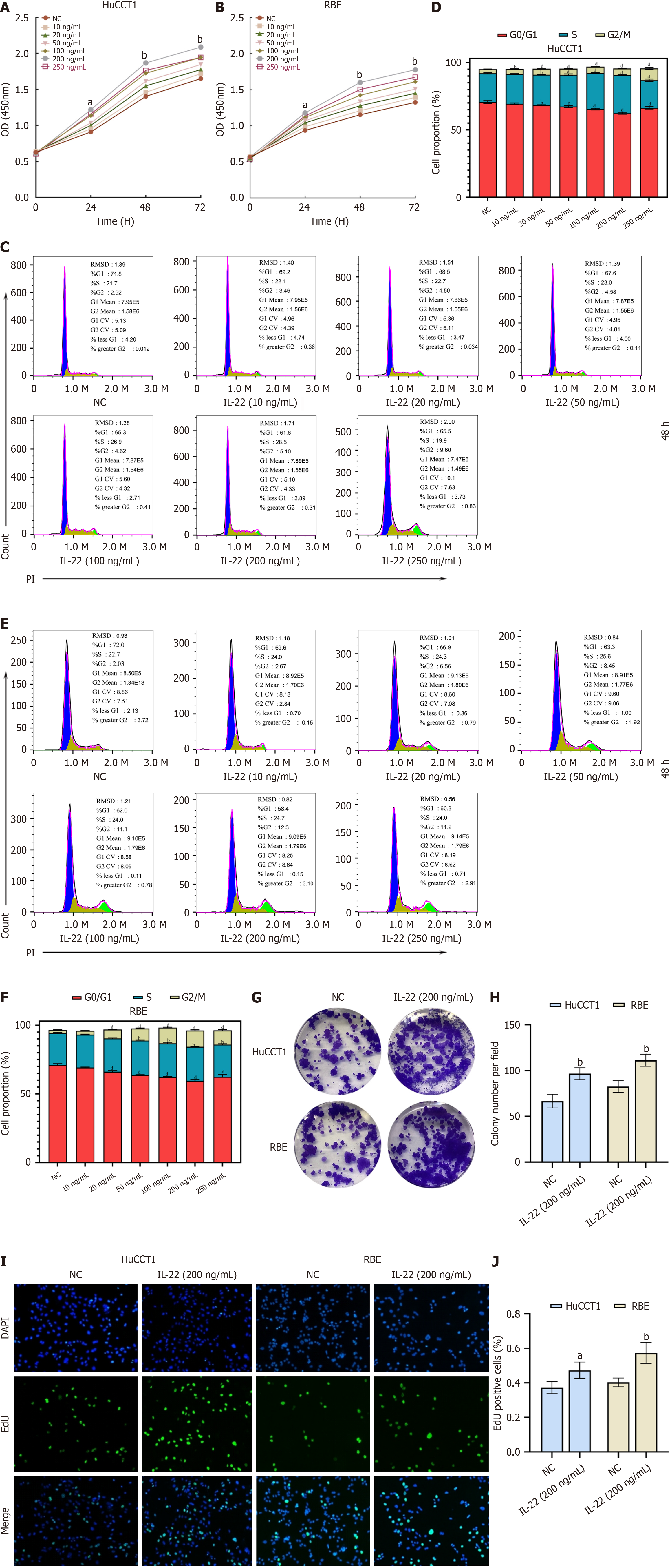
Figure 2 IL-22 promotes the proliferation of cholangiocarcinoma cells.
A: CCK8 proliferation curves of HuCCT1 cells stimulated with different concentrations of IL-22 at various time points; B: CCK8 proliferation curves of RBE cells stimulated with different concentrations of IL-22 at various time points; C: Flow cytometry cell cycle profiles of HuCCT1 cells after stimulation with different concentrations of IL-22 for 48 hours; D: Quantitative analysis results of the cell cycle assessment by flow cytometry; E: Flow cytometry cell cycle profiles of RBE cells after stimulation with different concentrations of IL-22 for 48 hours; F: Quantitative analysis results of the cell cycle assessment by flow cytometry; G and H: Colony formation and quantitative analysis results of HuCCT1 and RBE cells after stimulation with IL-22 at a concentration of 200 ng/mL; I and J: EdU incorporation and quantitative analysis results of HuCCT1 and RBE cells after stimulation with IL-22 at a concentration of 200 ng/mL. aP < 0.05, bP < 0.01. NC: Negative control.
- Citation: Zhou J, Chen JR, Li JM, Han SQ, Deng XY, Li ZM, Tong W, Wang C, Bai Y, Zhang YM. IL-22/IL-22R1 pathway enhances cholangiocarcinoma progression via ERK1/2 activation. World J Gastrointest Oncol 2025; 17(3): 102083
- URL: https://www.wjgnet.com/1948-5204/full/v17/i3/102083.htm
- DOI: https://dx.doi.org/10.4251/wjgo.v17.i3.102083


