Copyright
©The Author(s) 2025.
World J Gastrointest Oncol. Mar 15, 2025; 17(3): 102083
Published online Mar 15, 2025. doi: 10.4251/wjgo.v17.i3.102083
Published online Mar 15, 2025. doi: 10.4251/wjgo.v17.i3.102083
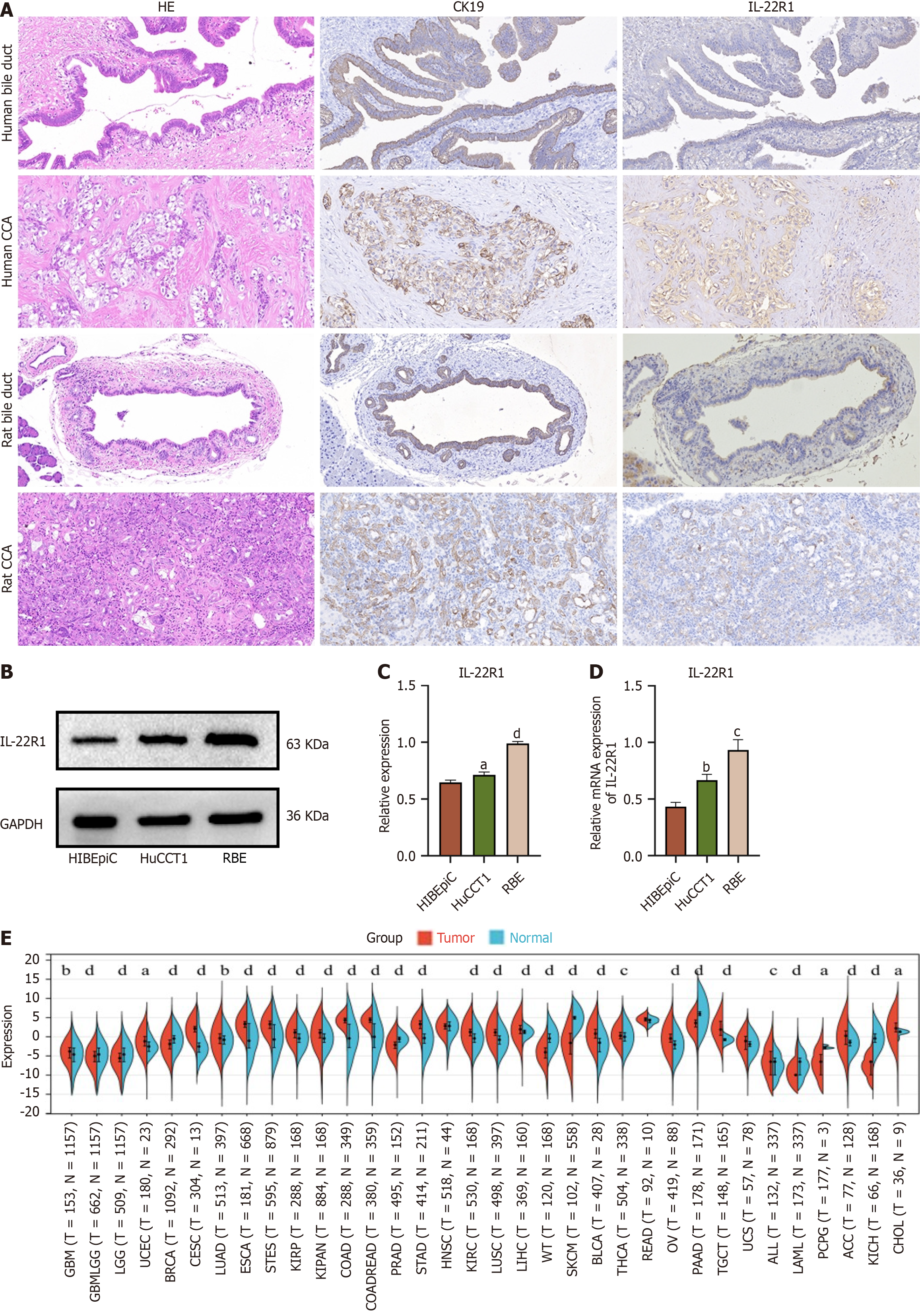
Figure 1 Aberrant expression of IL-22R1 in cholangiocarcinoma cell Lines, human and rat bile ducts, and cancer tissues.
A: Expression of IL-22R1 in human bile duct tissues and cholangiocarcinoma tissues (from left to right: HE staining, immunohistochemical results for CK19, immunohistochemical results for IL-22R1); Expression of IL-22R1 in rat bile duct tissues and cholangiocarcinoma tissues (TAA-induced; from left to right: HE staining, immunohistochemical results for CK19, immunohistochemical results for IL-22R1); B: Western blot analysis showing IL-22R1 expression in RBE, HuCCT1, and HIBEpiCs cells; C: Quantitative analysis of the relative expression of IL-22R1 in RBE, HuCCT1, and HIBEpiCs cells based on Western blot results. The expression in RBE and HuCCT1 is higher than that in HIBEpiCs cells; D: Quantitative analysis of the relative mRNA expression of IL-22R1 in RBE, HuCCT1, and HIBEpiCs cells. The expression in RBE and HuCCT1 is higher than that in HIBEpiCs cells; E: Expression of IL22R1 gene in various cancer samples extracted from the UCSC database. The expression in cholangiocarcinoma is higher than that in adjacent normal tissues. aP < 0.05, bP < 0.01, cP < 0.001, dP < 0.0001.
- Citation: Zhou J, Chen JR, Li JM, Han SQ, Deng XY, Li ZM, Tong W, Wang C, Bai Y, Zhang YM. IL-22/IL-22R1 pathway enhances cholangiocarcinoma progression via ERK1/2 activation. World J Gastrointest Oncol 2025; 17(3): 102083
- URL: https://www.wjgnet.com/1948-5204/full/v17/i3/102083.htm
- DOI: https://dx.doi.org/10.4251/wjgo.v17.i3.102083


