Copyright
©The Author(s) 2025.
World J Gastrointest Oncol. Mar 15, 2025; 17(3): 101174
Published online Mar 15, 2025. doi: 10.4251/wjgo.v17.i3.101174
Published online Mar 15, 2025. doi: 10.4251/wjgo.v17.i3.101174
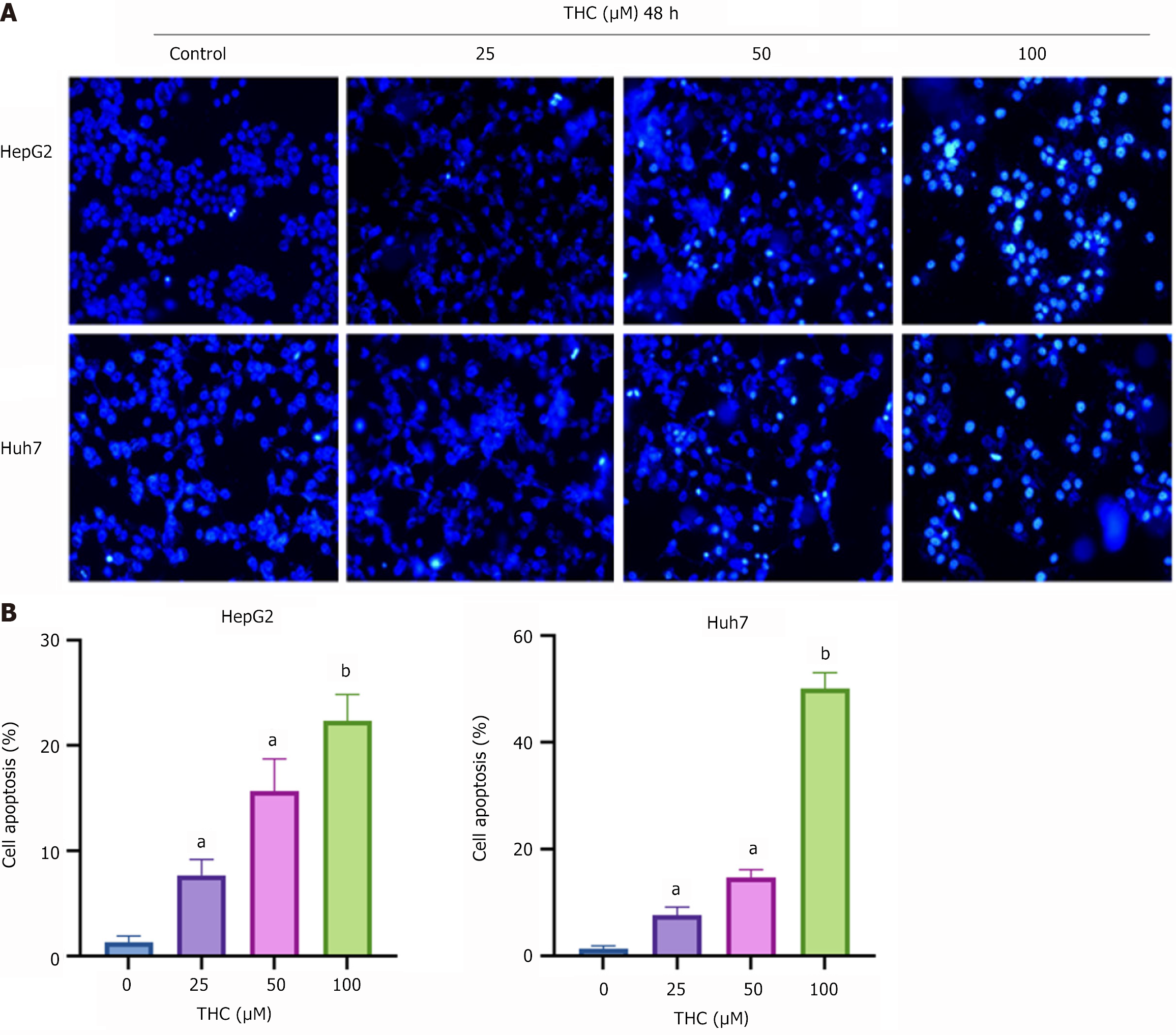
Figure 9 Tetrahydrocurcumin promoted the apoptosis of HepG2 and Huh7 cells.
A: Hoechst 33342 staining was used to detect the apoptosis of HepG2 and Huh7 cells treated with tetrahydrocurcumin (25, 50, or 100 μmol/L) for 48 hours, after photographing under a fluorescence microscope (20 µm); B: Graphs of apoptosis rates of HepG2 and Huh7 cells determined using Hoechst 33342 staining. All data are presented as mean ± SD, n = 3. Compared to the control group, aP < 0.05; bP < 0.01; THC: Tetrahydrocurcumin.
- Citation: Bao ZC, Zhang Y, Liu ZD, Dai HJ, Ren F, Li N, Lv SY, Zhang Y. Tetrahydrocurcumin-induced apoptosis of hepatocellular carcinoma cells involves the TP53 signaling pathway, as determined by network pharmacology. World J Gastrointest Oncol 2025; 17(3): 101174
- URL: https://www.wjgnet.com/1948-5204/full/v17/i3/101174.htm
- DOI: https://dx.doi.org/10.4251/wjgo.v17.i3.101174


