Copyright
©The Author(s) 2025.
World J Gastrointest Oncol. Mar 15, 2025; 17(3): 100861
Published online Mar 15, 2025. doi: 10.4251/wjgo.v17.i3.100861
Published online Mar 15, 2025. doi: 10.4251/wjgo.v17.i3.100861
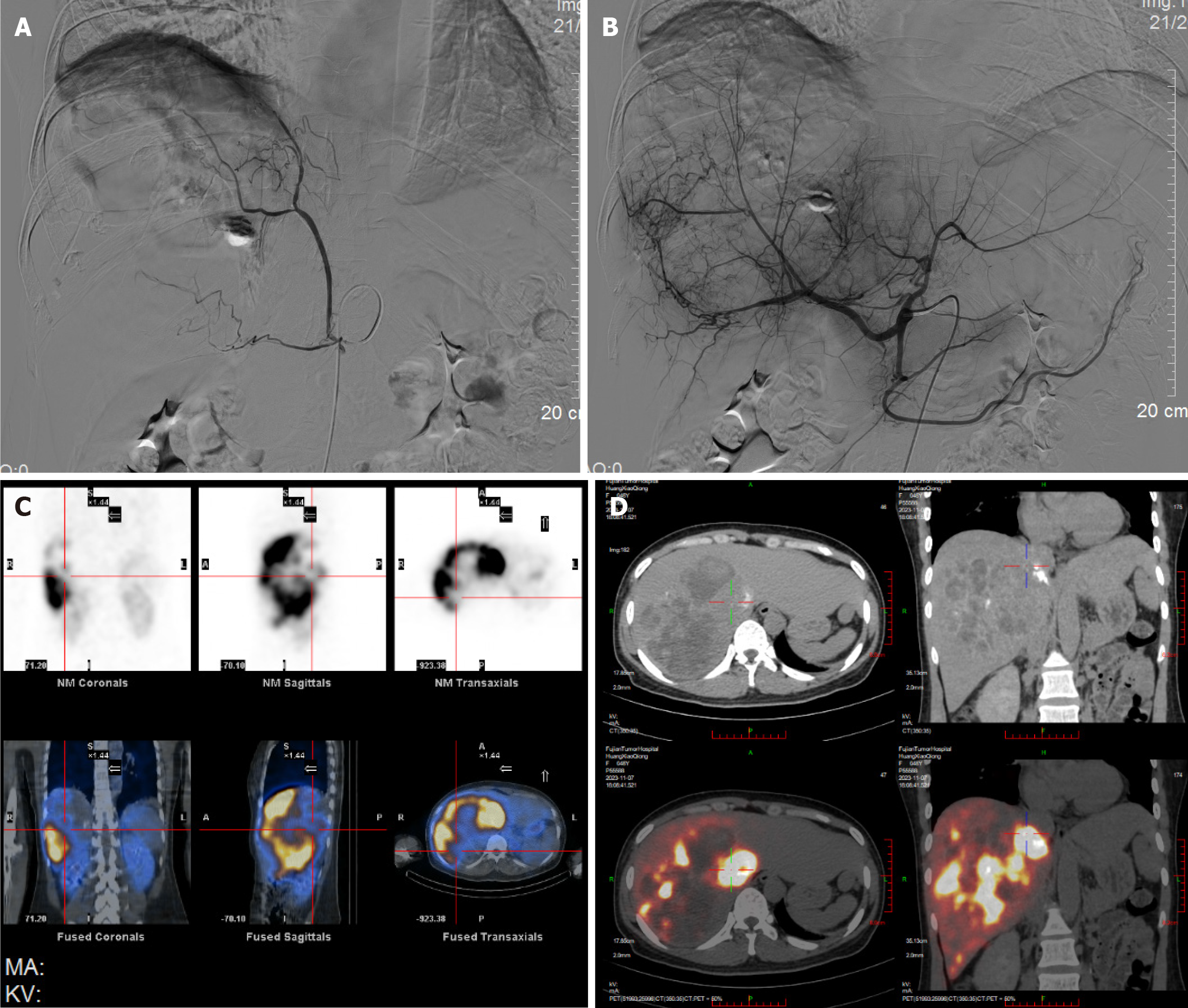
Figure 2 Treatment.
A: The angiography revealed that the right diaphragmatic artery emitted an aberrant vessel, which was involved in some tumor blood supply; B: The angiography revealed that the main blood supply artery of the tumor came from the right hepatic artery; C: The lung shunting fraction was 12.68%, with a T/N of 4.28. The partition model revealed that the prescription activity was 3.8 GBq at a tumoral dose of 110 Gy; D: Single-photon emission computed tomography/computed tomography at 1 hour after selective internal radiation therapy showed that the right lobe of liver mass was covered with yttrium-90 resin microspheres. There is no significant shunt in the extrahepatic tissues within the field of vision. Multiple enlarged lymph nodes in the hepatic hilum and retroperitoneum. Right oblique fissure pleural calcification, slight chronic inflammation in both lungs, and slight pleural effusion in the right chest.
- Citation: Hao MZ, Lin HL, Hu YB, Chen QZ, Chen ZX, Qiu LB, Lin DY, Zhang H, Zheng DC, Fang ZT, Liu JF. Combination therapy strategy based on selective internal radiation therapy as conversion therapy for inoperable giant hepatocellular carcinoma: A case report. World J Gastrointest Oncol 2025; 17(3): 100861
- URL: https://www.wjgnet.com/1948-5204/full/v17/i3/100861.htm
- DOI: https://dx.doi.org/10.4251/wjgo.v17.i3.100861


