Copyright
©The Author(s) 2025.
World J Gastrointest Oncol. Mar 15, 2025; 17(3): 100861
Published online Mar 15, 2025. doi: 10.4251/wjgo.v17.i3.100861
Published online Mar 15, 2025. doi: 10.4251/wjgo.v17.i3.100861
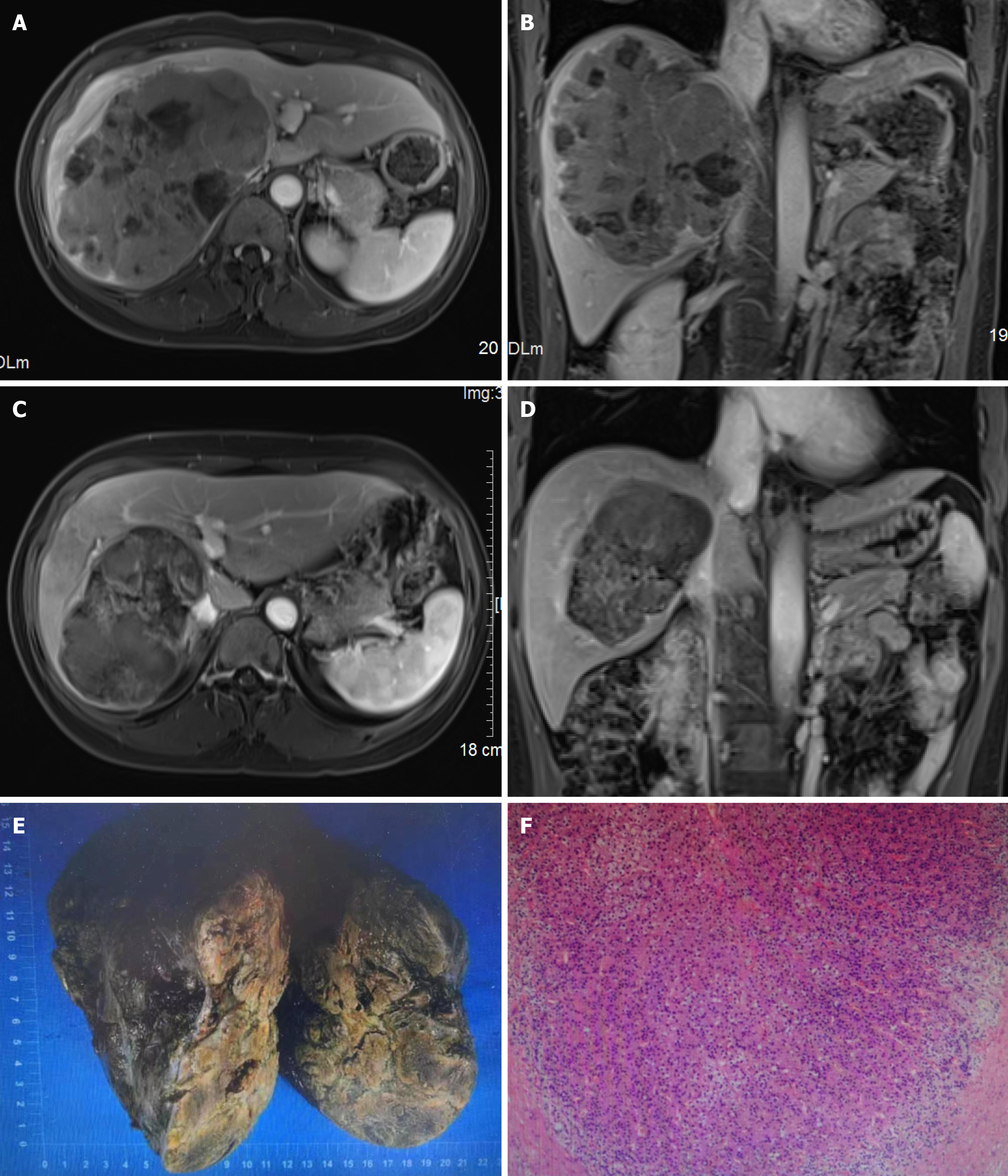
Figure 1 Imaging examinations.
A and B: Enhanced magnetic resonance imaging on 24 October 2023, showed the giant tumor in size of 15.8 cm × 11.4 cm × 14.8 cm, accompanied by the invasion of the right portal vein as well as right and middle hepatic veins; C and D: Enhanced magnetic resonance imaging on 28 February 2024, showed a tumor size of 11.6 cm × 7.7 cm × 10.1 cm, indicating a complete response according to mRECIST; E and F: The hepatic tissue size was 17 cm × 14 cm × 10 cm. Upon incision, a grayish yellow and grayish white mass with a size of 13.5 cm × 12 cm × 9.5 cm was observed as a nodular gray-white and grayish red mass with extensive necrosis.
- Citation: Hao MZ, Lin HL, Hu YB, Chen QZ, Chen ZX, Qiu LB, Lin DY, Zhang H, Zheng DC, Fang ZT, Liu JF. Combination therapy strategy based on selective internal radiation therapy as conversion therapy for inoperable giant hepatocellular carcinoma: A case report. World J Gastrointest Oncol 2025; 17(3): 100861
- URL: https://www.wjgnet.com/1948-5204/full/v17/i3/100861.htm
- DOI: https://dx.doi.org/10.4251/wjgo.v17.i3.100861


