Copyright
©The Author(s) 2025.
World J Gastrointest Oncol. Feb 15, 2025; 17(2): 98808
Published online Feb 15, 2025. doi: 10.4251/wjgo.v17.i2.98808
Published online Feb 15, 2025. doi: 10.4251/wjgo.v17.i2.98808
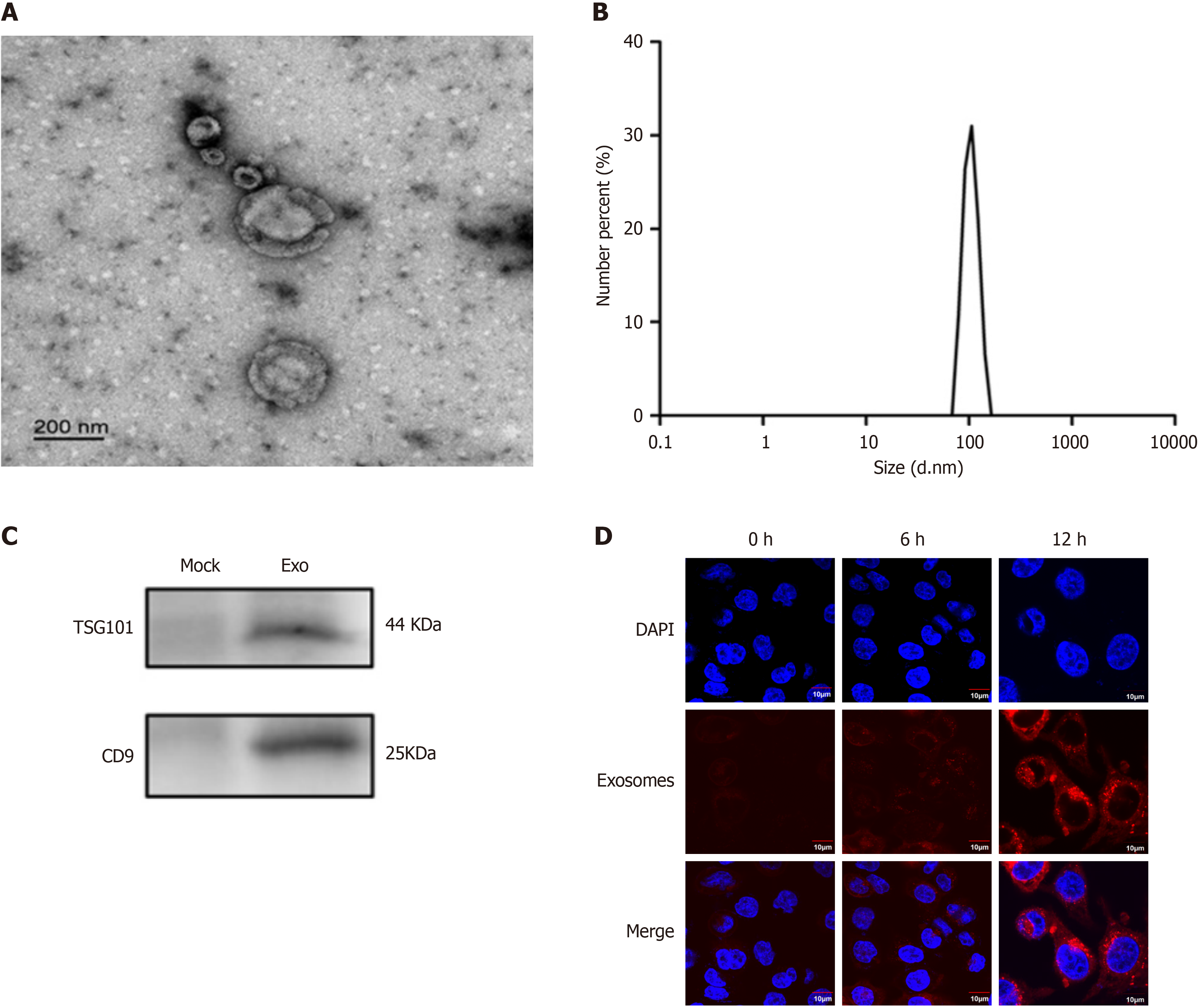
Figure 3 Characterization of extracted exosomes.
A: Transmission electron microscopy image of exosomes (scale bar = 100 nm); B: Nanoparticle tracking analysis of exosome concentration and size; C: Western blot detection of exosomal-specific markers TSG101 and CD9; D: Uptake of exosomes by KYSE30 cells.
- Citation: Ge YY, Xia XC, Wu AQ, Ma CY, Yu LH, Zhou JY. Identifying adipocyte-derived exosomal miRNAs as potential novel prognostic markers for radiotherapy of esophageal squamous cell carcinoma. World J Gastrointest Oncol 2025; 17(2): 98808
- URL: https://www.wjgnet.com/1948-5204/full/v17/i2/98808.htm
- DOI: https://dx.doi.org/10.4251/wjgo.v17.i2.98808


