Copyright
©The Author(s) 2025.
World J Gastrointest Oncol. Jan 15, 2025; 17(1): 100757
Published online Jan 15, 2025. doi: 10.4251/wjgo.v17.i1.100757
Published online Jan 15, 2025. doi: 10.4251/wjgo.v17.i1.100757
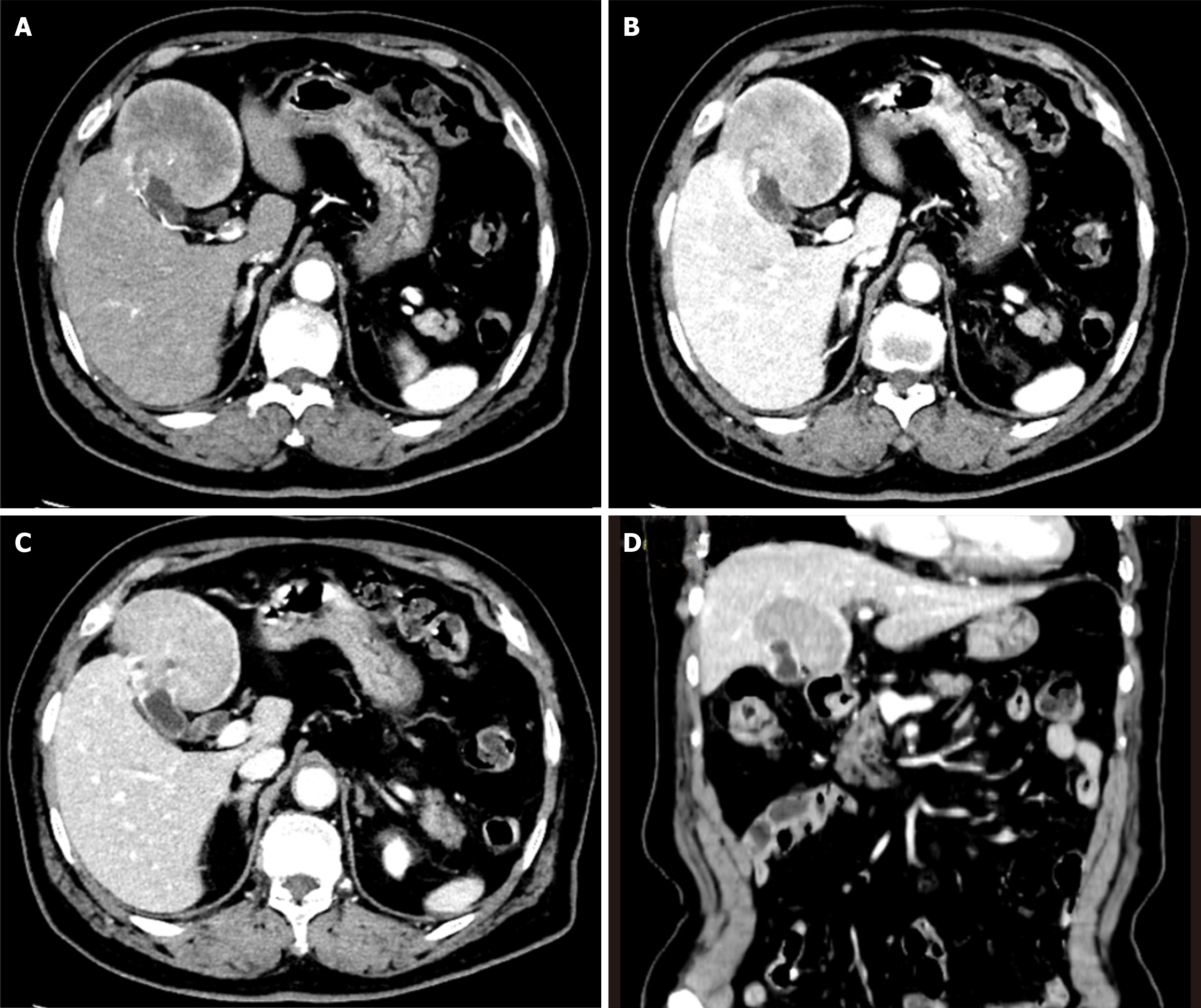
Figure 1 Abdominal contrast-enhanced computed tomography scan reveals a 7 cm mass in liver segments IVb and V, with localized thickening of the gallbladder fundus wall and early-stage enhancement followed by a prolonged contrast effect.
A: Arterial phase showed inhomogeneous lesion enhancement; B: The portal venous phase showed a prolonged contrast effect in the mass; C: The delayed phase showed progressive enhancement of the mass; D: The mass invades liver segments IVb and V.
- Citation: Yang YC, Chen ZT, Wan DL, Tang H, Liu ML. Targeted gene sequencing and bioinformatics analysis of patients with gallbladder neuroendocrine carcinoma: A case report. World J Gastrointest Oncol 2025; 17(1): 100757
- URL: https://www.wjgnet.com/1948-5204/full/v17/i1/100757.htm
- DOI: https://dx.doi.org/10.4251/wjgo.v17.i1.100757


