Copyright
©The Author(s) 2024.
World J Gastrointest Oncol. Sep 15, 2024; 16(9): 4028-4036
Published online Sep 15, 2024. doi: 10.4251/wjgo.v16.i9.4028
Published online Sep 15, 2024. doi: 10.4251/wjgo.v16.i9.4028
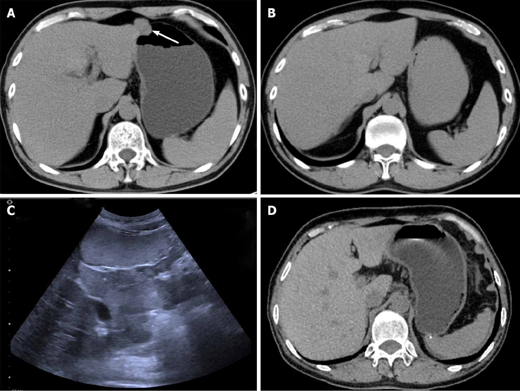
Figure 6 Postoperative follow-up examination results in September 2023.
Comparison of pre- and postoperative computed tomography (CT) images of the male patient. A: Preoperative CT plain scan of the whole abdomen with localized nodular shadows in the antrum of the stomach; B: Postoperative CT plain scan of the whole abdomen with postoperative changes in the distal stomach and no abnormal signs in the anastomosis; C: Gastrointestinal ultrasonography showing postoperative gastrointestinal stromal tumor, with no space-occupying lesions inside or outside the gastric lumen; D: Postoperative CT of the female patient showing postoperative changes after partial resection of the gastric fundus with no abnormal signs in the anastomosis.
- Citation: Wang XK, Shen LF, Yang X, Su H, Wu T, Tao PX, Lv HY, Yao TH, Yi L, Gu YH. Two different mutational types of familial gastrointestinal stromal tumors: Two case reports. World J Gastrointest Oncol 2024; 16(9): 4028-4036
- URL: https://www.wjgnet.com/1948-5204/full/v16/i9/4028.htm
- DOI: https://dx.doi.org/10.4251/wjgo.v16.i9.4028


