Copyright
©The Author(s) 2024.
World J Gastrointest Oncol. Sep 15, 2024; 16(9): 4006-4013
Published online Sep 15, 2024. doi: 10.4251/wjgo.v16.i9.4006
Published online Sep 15, 2024. doi: 10.4251/wjgo.v16.i9.4006
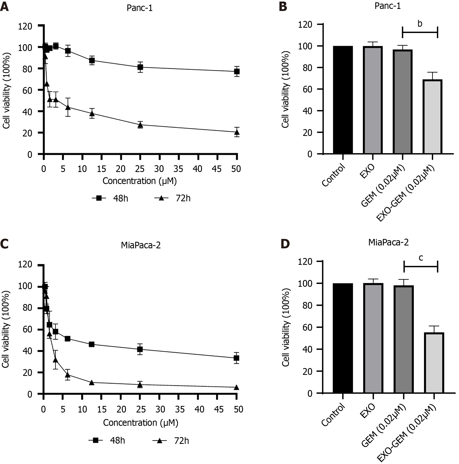
Figure 2 The cytotoxicity of free gemcitabine and exosomes loaded with gemcitabine against Panc-1 and MiaPaca-2 cells.
A-D: The cells were treated in triplicate with the indicated concentrations of gemcitabine, exosomes loaded with gemcitabine, or exosomes for 48 or 72 hours. The viability of each group of cells was assessed by the MTT assay. The cyttoxicity of different concentrations of gemcitabine against Panc-1 (A) and MiaPaca-2 cells (C); comparison of effects of gemcitabine, exosomes loaded with gemcitabine and control exosomes on the viability of Panc-1 (B) and MiaPaca-2 (D) cells. Data are represented as the mean ± SD (n = 3) of a representative experiment. bP < 0.01, vs the gemcitabine group; cP < 0.001, vs the gemcitabine group. EXO: Exosomes; GEM: Gemcitabine; EXO-GEM: Exosomes loaded with gemcitabine.
- Citation: Tang ZG, Chen TM, Lu Y, Wang Z, Wang XC, Kong Y. Human bone marrow mesenchymal stem cell-derived exosomes loaded with gemcitabine inhibit pancreatic cancer cell proliferation by enhancing apoptosis. World J Gastrointest Oncol 2024; 16(9): 4006-4013
- URL: https://www.wjgnet.com/1948-5204/full/v16/i9/4006.htm
- DOI: https://dx.doi.org/10.4251/wjgo.v16.i9.4006


