Copyright
©The Author(s) 2024.
World J Gastrointest Oncol. Aug 15, 2024; 16(8): 3723-3731
Published online Aug 15, 2024. doi: 10.4251/wjgo.v16.i8.3723
Published online Aug 15, 2024. doi: 10.4251/wjgo.v16.i8.3723
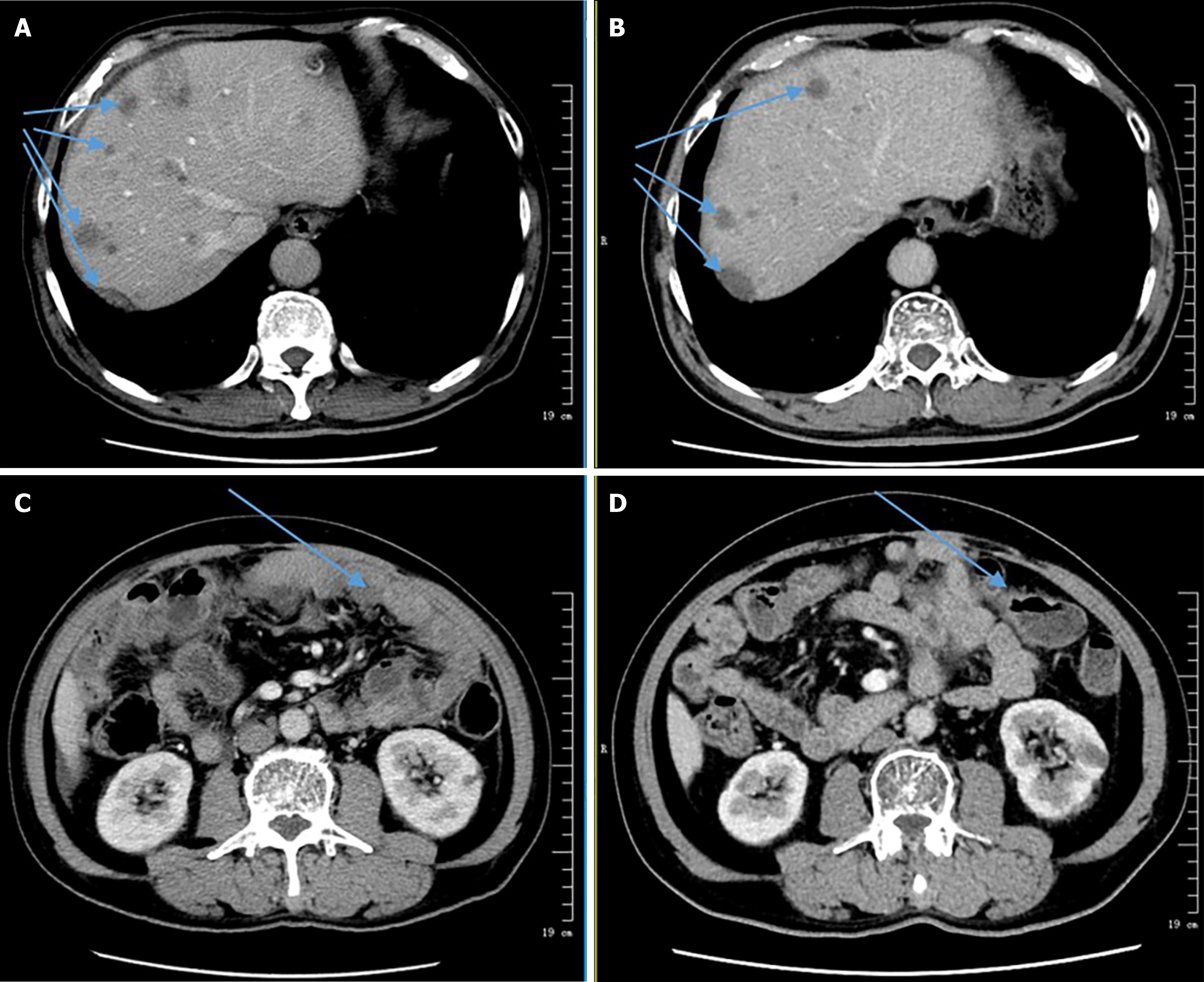
Figure 4 Comparison of Computed tomography.
A: Computed tomography (CT) images from May 18, 2022; B: CT images from September 14, 2022; A and B: The total number of liver lesions has decreased, and some lesions have reduced in enhancement compared to the previous images (indicated by the arrow); C: CT images from May 18, 2022; D: CT images from September 14, 2022; C and D: Showing decreased peritoneal thickening, reduced omentum opacity, and decreased size of some nodules in the abdominal cavity. There is also a reduction in abdominal and pelvic fluid accumulation (indicated by the arrow).
- Citation: Feng Q, Yu W, Feng JH, Huang Q, Xiao GX. Jejunal sarcomatoid carcinoma: A case report and review of literature. World J Gastrointest Oncol 2024; 16(8): 3723-3731
- URL: https://www.wjgnet.com/1948-5204/full/v16/i8/3723.htm
- DOI: https://dx.doi.org/10.4251/wjgo.v16.i8.3723


