Copyright
©The Author(s) 2024.
World J Gastrointest Oncol. Aug 15, 2024; 16(8): 3723-3731
Published online Aug 15, 2024. doi: 10.4251/wjgo.v16.i8.3723
Published online Aug 15, 2024. doi: 10.4251/wjgo.v16.i8.3723
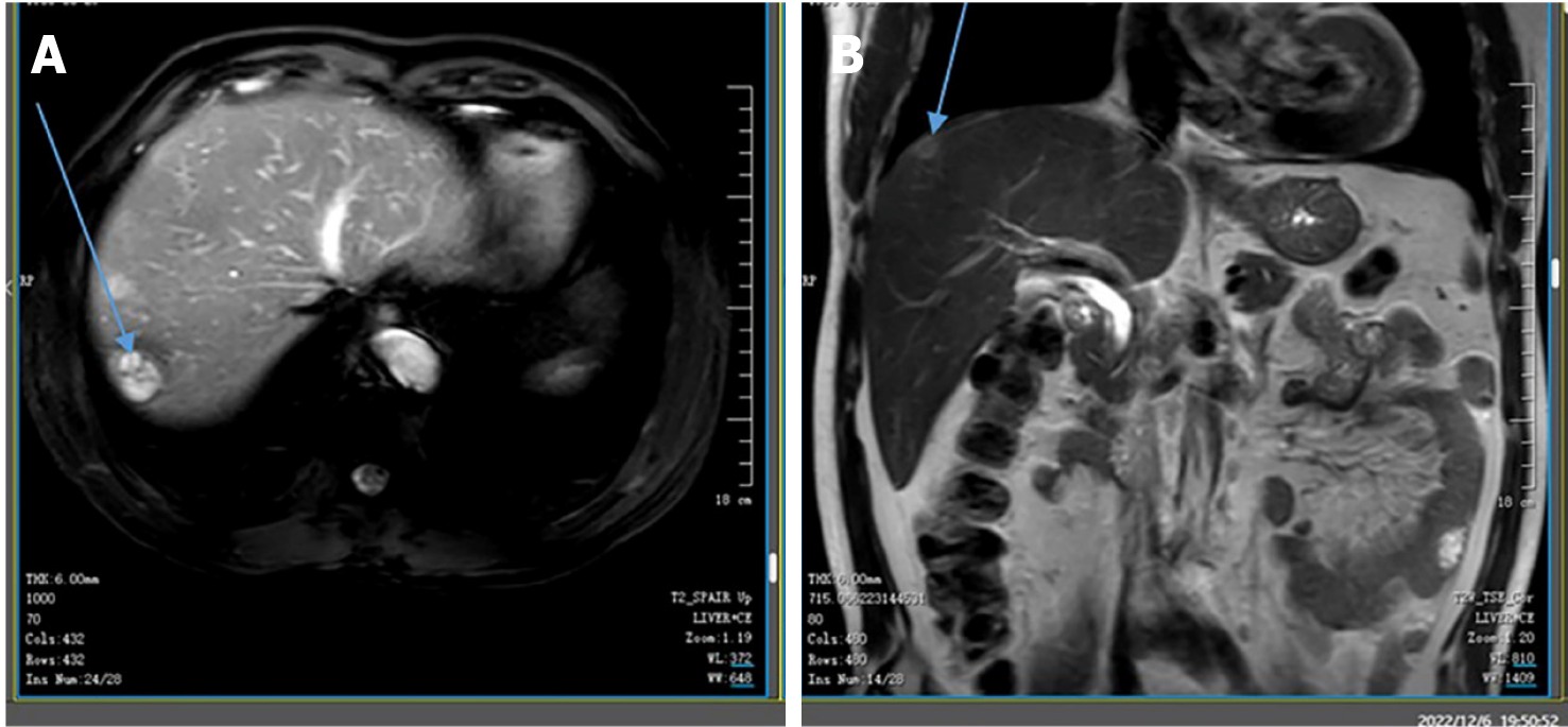
Figure 1 Liver magnetic resonance images.
Magnetic resonance images performed at Yichang Central People's Hospital on February 9, 2022. Well-circumscribed, heterogeneous, and hypodense masses are located in the right posterior lobe of the liver, measuring 2.1 cm × 2.0 cm (indicated by the arrow). A: Transverse view; B: Coronal view.
- Citation: Feng Q, Yu W, Feng JH, Huang Q, Xiao GX. Jejunal sarcomatoid carcinoma: A case report and review of literature. World J Gastrointest Oncol 2024; 16(8): 3723-3731
- URL: https://www.wjgnet.com/1948-5204/full/v16/i8/3723.htm
- DOI: https://dx.doi.org/10.4251/wjgo.v16.i8.3723


