Copyright
©The Author(s) 2024.
World J Gastrointest Oncol. Aug 15, 2024; 16(8): 3716-3722
Published online Aug 15, 2024. doi: 10.4251/wjgo.v16.i8.3716
Published online Aug 15, 2024. doi: 10.4251/wjgo.v16.i8.3716
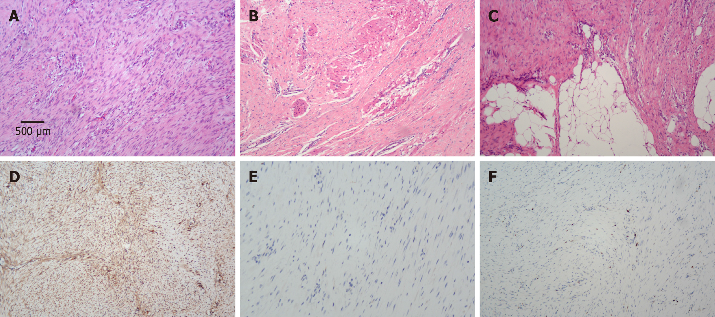
Figure 3 Pathological and immunohistochemical findings.
A-C: Hematoxylin-eosin staining showed that the tumor cells displayed a fascicular or interwoven pattern (A) and infiltrated into the muscularis propria of the sigmoid colon (B) and the adipose tissue (C) (× 100); D-F: Representative images of immunohistochemical staining for β-catenin (D), CD117 (E) and Ki-67 (F) in tumor cells (× 100).
- Citation: Yu PP, Liu XC, Yin L, Yin G. Aggressive fibromatosis of the sigmoid colon: A case report. World J Gastrointest Oncol 2024; 16(8): 3716-3722
- URL: https://www.wjgnet.com/1948-5204/full/v16/i8/3716.htm
- DOI: https://dx.doi.org/10.4251/wjgo.v16.i8.3716


