Copyright
©The Author(s) 2024.
World J Gastrointest Oncol. Aug 15, 2024; 16(8): 3651-3671
Published online Aug 15, 2024. doi: 10.4251/wjgo.v16.i8.3651
Published online Aug 15, 2024. doi: 10.4251/wjgo.v16.i8.3651
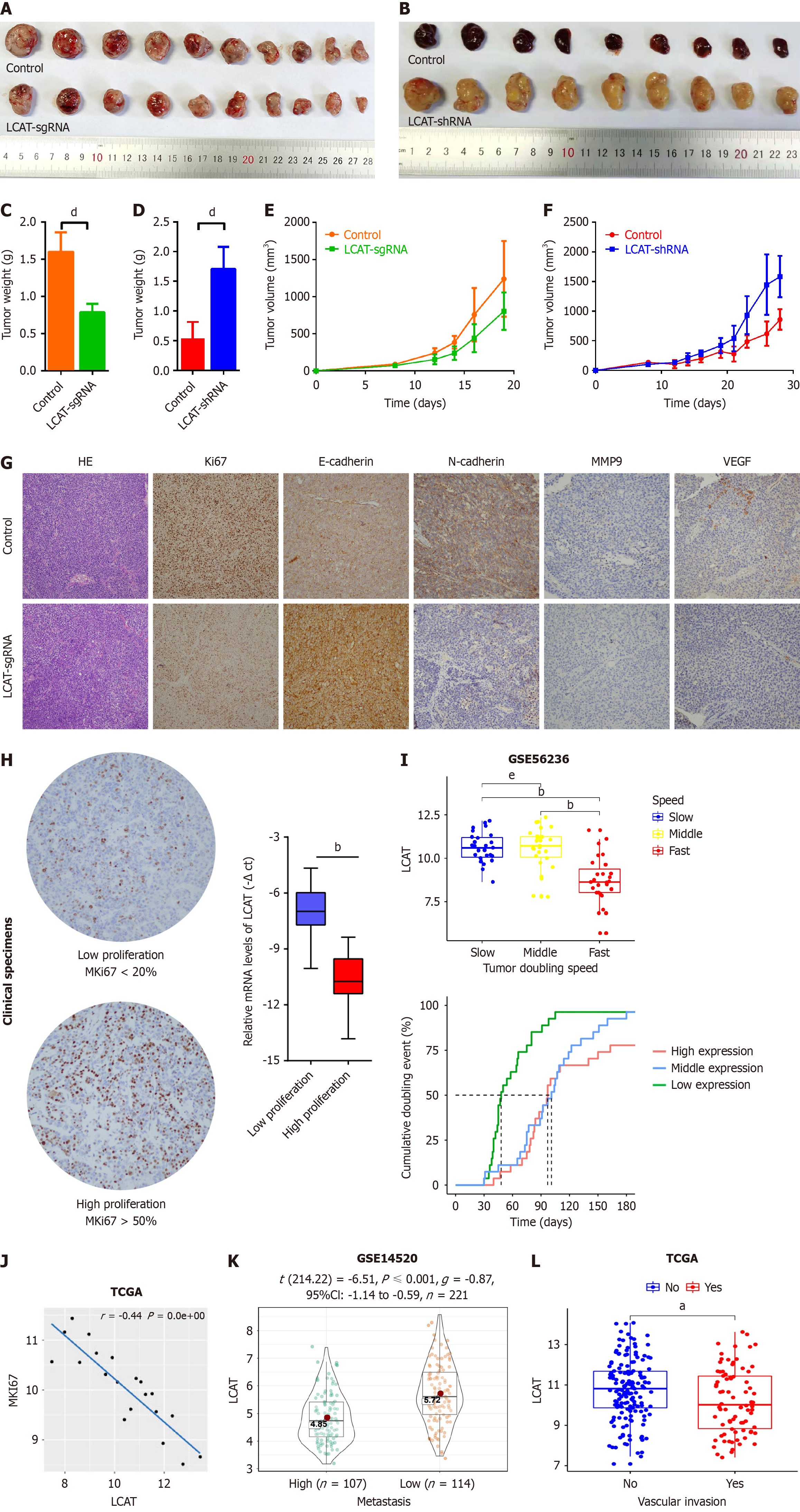
Figure 3 Lecithin-cholesterol acyltransferase inhibits tumor growth and metastasis potential in vivo and is negatively correlated with tumor growth, metastasis and vascular invasion of hepatocellular carcinoma patients.
A-F: Huh7 and lecithin-cholesterol acyltransferase (LCAT) overexpressed Huh7 or HepG2 and LCAT knocked down HepG2 cells were used to perform the tumor formation assay in nude mice. The tumors were dissected out on day 28 (A and B). The volume (C and D) and weight (E and F) of xenograft tumors were measured at indicated time points; G: Sections of xenograft tumors were stained with hematoxylin and eosin, MKI-67, E-cadherin, N-cadherin, matrix metalloproteinase 9 (MMP9), and vascular endothelial growth factor (VEGF); H: The sections of hepatocellular carcinoma (HCC) tissues were stained with MKI67. The high and low proliferative of HCC tissues were defined by MKI67 over 50% and MKI67 lower than 20% respectively (left). The mRNA levels of LCAT in HCC tissues were quantified by quantitative real-time polymerase chain reaction (right); I: Expression levels of LCAT in different growth speeds and the relationship between LCAT expression levels and tumor doubling time in the GSE54236 dataset were analyzed; J: Correlation analysis of LCAT mRNA expression with MKI67 using the The Cancer Genome Atlas (TCGA)-LICH dataset; K: Decreased LCAT exhibited in high metastasis HCC patients according to Wang’s cohort (GSE14520); L: Decreased LCAT exhibited in HCC patients with vascular invasion according to TCGA-LICH cohort. aP < 0.01, bP < 0.0001, dP < 0.001, eNS: Not significant. LCAT: Lecithin-cholesterol acyltransferase; TCGA: The Cancer Genome Atlas.
- Citation: Li Y, Jiang LN, Zhao BK, Li ML, Jiang YY, Liu YS, Liu SH, Zhu L, Ye X, Zhao JM. Lecithin-cholesterol acyltransferase is a potential tumor suppressor and predictive marker for hepatocellular carcinoma metastasis. World J Gastrointest Oncol 2024; 16(8): 3651-3671
- URL: https://www.wjgnet.com/1948-5204/full/v16/i8/3651.htm
- DOI: https://dx.doi.org/10.4251/wjgo.v16.i8.3651


