Copyright
©The Author(s) 2024.
World J Gastrointest Oncol. Aug 15, 2024; 16(8): 3471-3480
Published online Aug 15, 2024. doi: 10.4251/wjgo.v16.i8.3471
Published online Aug 15, 2024. doi: 10.4251/wjgo.v16.i8.3471
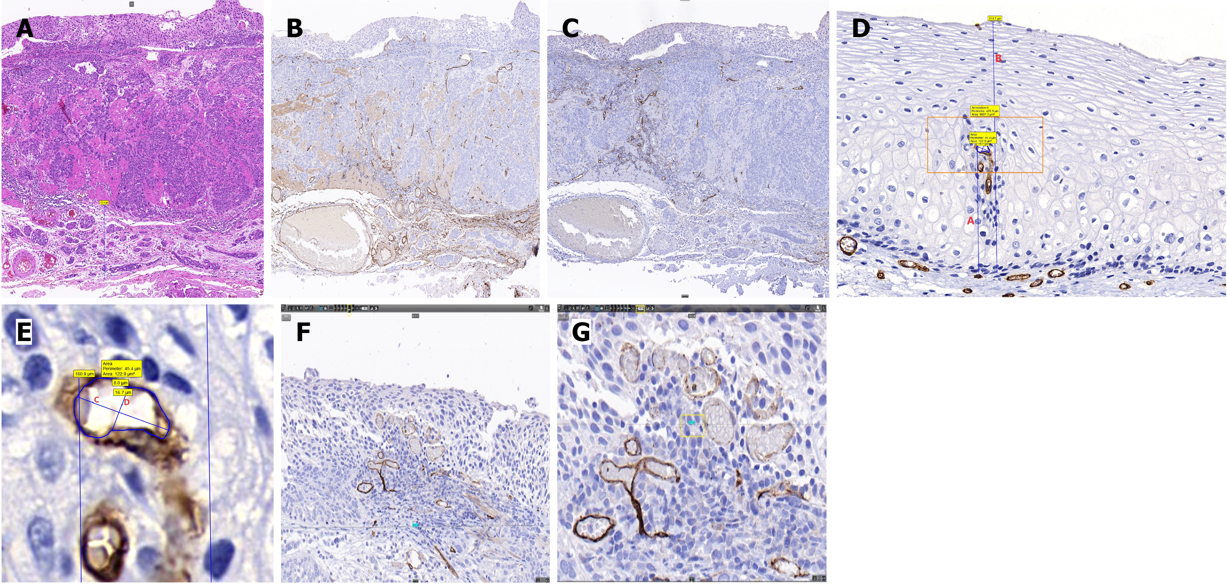
Figure 2 Pathological data collection using Case Viewer software.
A: Pathological slide stained with hematoxylin and eosin (magnification: × 100); B: Pathological slide stained for CD34 (magnification: × 100); C: Pathological slide stained for D2-40 (magnification: × 100); D: Measurement of intrapapillary capillary loop (IPCL)-related characteristics (red A: Apical distance from the vertex of the IPCL to the basement membrane; red B: Thickness of the epithelial layer); E: Enlarged orange region in picture D (red C: Short caliber of the IPCL; red D: Long caliber of the IPCL; and the region enclosed by the blue line is the area of the IPCL); F: Pathological slide stained for CD34 (magnification: × 800); the transverse diameter of each field of view was 719.8 μm; G: Pathological slide stained for CD34 (magnification: × 2000); the transverse diameter of each field of view is 287.9 μm.
- Citation: Shu WY, Shi YY, Huang JT, Meng LM, Zhang HJ, Cui RL, Li Y, Ding SG. Microvascular structural changes in esophageal squamous cell carcinoma pathology according to intrapapillary capillary loop types under magnifying endoscopy. World J Gastrointest Oncol 2024; 16(8): 3471-3480
- URL: https://www.wjgnet.com/1948-5204/full/v16/i8/3471.htm
- DOI: https://dx.doi.org/10.4251/wjgo.v16.i8.3471


