Copyright
©The Author(s) 2024.
World J Gastrointest Oncol. Aug 15, 2024; 16(8): 3471-3480
Published online Aug 15, 2024. doi: 10.4251/wjgo.v16.i8.3471
Published online Aug 15, 2024. doi: 10.4251/wjgo.v16.i8.3471
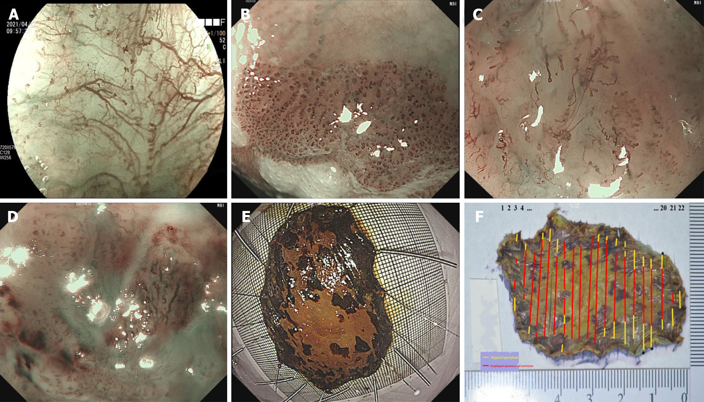
Figure 1 Intrapapillary capillary loops in superficial esophageal lesions under magnifying endoscopy and photographic recordings of both fresh and fixed specimens.
A: Intrapapillary capillary loops (IPCLs) of type A under narrow-band imaging or blue laser imaging combined with magnifying endoscopy (ME-NBI/BLI) (magnification: × 80); B: IPCLs of type B1 under ME-NBI/BLI (magnification: × 80); C: IPCLs of type B2 under ME-NBI/BLI (magnification: × 80); D: IPCLs of type B3 under ME-NBI/BLI (magnification: × 80); E: Flesh specimen after endoscopic resection; F: Pathologically fixed specimen with the red line marking the extent of the cancer and the yellow line marking the extent of intraepithelial neoplasia.
- Citation: Shu WY, Shi YY, Huang JT, Meng LM, Zhang HJ, Cui RL, Li Y, Ding SG. Microvascular structural changes in esophageal squamous cell carcinoma pathology according to intrapapillary capillary loop types under magnifying endoscopy. World J Gastrointest Oncol 2024; 16(8): 3471-3480
- URL: https://www.wjgnet.com/1948-5204/full/v16/i8/3471.htm
- DOI: https://dx.doi.org/10.4251/wjgo.v16.i8.3471


