Copyright
©The Author(s) 2024.
World J Gastrointest Oncol. Jul 15, 2024; 16(7): 3350-3356
Published online Jul 15, 2024. doi: 10.4251/wjgo.v16.i7.3350
Published online Jul 15, 2024. doi: 10.4251/wjgo.v16.i7.3350
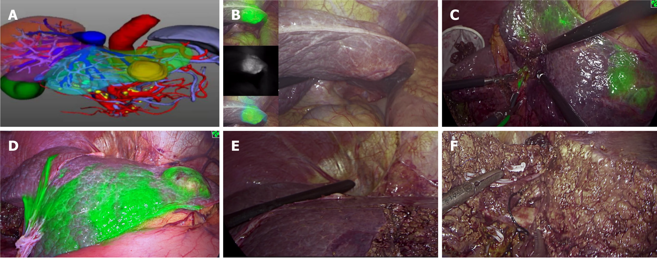
Figure 4 Intraoperative images during the first operation.
A: Preoperative three-dimensional reconstruction for the watershed analysis; B: Different fluorescence modes of liver lesions were visualized, and micrometastases in the liver could be screened simultaneously; C: Indocyanine green was injected into the portal watershed of the S3 segment, and the S3 segment was stained; D: The tumor boundary and boundaries of the S3 and S2 segments were clearly visible; E: Intra-operative ultrasonography was used to investigate the remaining part of the liver. Simultaneously, the location and course of the left hepatic vein were investigated with the aid of ultrasonography, which ensured that the left hepatic vein was intact and exposed; F: Cross-section of the liver after resection of segment S2. The morphology and course of the left hepatic vein could be clearly seen.
- Citation: Wan DD, Li XJ, Wang XR, Liu TX. Metachronous multifocal carcinoma: A case report. World J Gastrointest Oncol 2024; 16(7): 3350-3356
- URL: https://www.wjgnet.com/1948-5204/full/v16/i7/3350.htm
- DOI: https://dx.doi.org/10.4251/wjgo.v16.i7.3350


