Copyright
©The Author(s) 2024.
World J Gastrointest Oncol. Jul 15, 2024; 16(7): 3350-3356
Published online Jul 15, 2024. doi: 10.4251/wjgo.v16.i7.3350
Published online Jul 15, 2024. doi: 10.4251/wjgo.v16.i7.3350
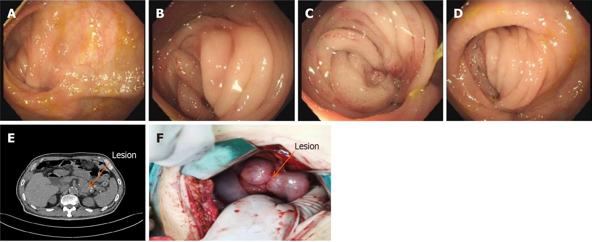
Figure 3 Schematic diagram of the colonoscopic and abdominal computed tomography results before the second operation and the lesion during the operation.
A-D: Entering the scope 70 cm from the anus. Anastomotic changes were visible. There was mucosal congestion and entanglement, and it was difficult to enter the scope; E: Dilatation and pneumatization of the bowel in the upper abdomen. When wide and large air-fluid planes could be seen, intestinal obstruction was considered. When the original lesion in the tail of the pancreas was enlarged compared with the previous one with unclear borders, cystadenoma was considered; F: Intra-operatively, the original anastomosis was found to be entangled and narrowed, and the posterior pancreatic lesion invaded the original anastomosis, which was confirmed by pathological examination.
- Citation: Wan DD, Li XJ, Wang XR, Liu TX. Metachronous multifocal carcinoma: A case report. World J Gastrointest Oncol 2024; 16(7): 3350-3356
- URL: https://www.wjgnet.com/1948-5204/full/v16/i7/3350.htm
- DOI: https://dx.doi.org/10.4251/wjgo.v16.i7.3350


