Copyright
©The Author(s) 2024.
World J Gastrointest Oncol. Jul 15, 2024; 16(7): 3350-3356
Published online Jul 15, 2024. doi: 10.4251/wjgo.v16.i7.3350
Published online Jul 15, 2024. doi: 10.4251/wjgo.v16.i7.3350
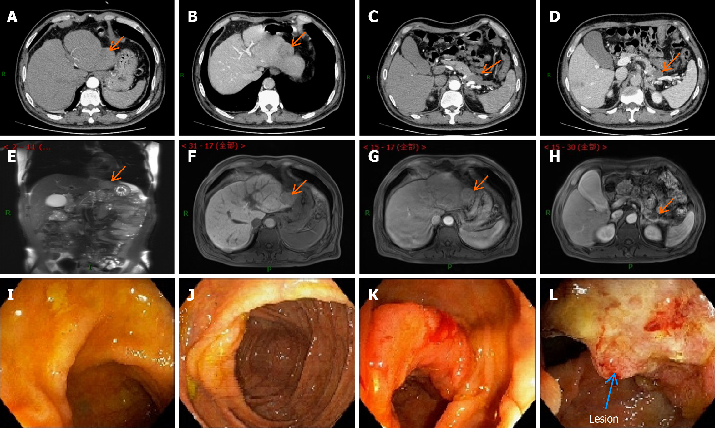
Figure 1 Schematic diagram of the enhanced computed tomography, magnetic resonance imaging, and colonoscopy results before the first surgery.
A: Contrast-enhanced abdominal computed tomography (CT) image of the liver during the arterial phase; B: Contrast-enhanced abdominal CT image of the liver during the portal phase; C: Contrast-enhanced abdominal CT image of the arterial pancreatic lesion; D: Contrast-enhanced abdominal CT image of the arterial pancreatic lesion; E-H: Contrast-enhanced magnetic resonance imaging (MRI) images of the liver and pancreatic foci at different levels. They suggest that the hepatic fissure was widened, and the edge of the liver was not smooth. The left lobe could be seen as a nodular abnormal signal shadow of about 35 mm × 31 mm in size. There was a low signal in T1-weighted image (T1WI) and a high signal in T2WI. There was mild enhancement in the arterial phase after enhancement, which receded in the delayed phase and was considered to be hepatocellular carcinoma. Pancreatic atrophy and the caudal part of the pancreas could be seen as cystic foci of about 25 mm × 28 mm in size. There was a low signal in T1WI and a high signal in T2WI. There was no enhancement in the arterial phase after enhancement, which was considered a cystic adenoma; I: Electron enteroscopic image of the end of the ileum; J: Electron enteroscopic image of the cecum; K: Electron enteroscopic image of the liver region; L: Electron enteroscopic image of the liver region, suggesting neoplasm in the liver region of the colon, a longitudinal change of 3 cm, and narrowing of the intestinal lumen by half. The results of the sampling pathology suggested moderately differentiated adenocarcinoma of the colon.
- Citation: Wan DD, Li XJ, Wang XR, Liu TX. Metachronous multifocal carcinoma: A case report. World J Gastrointest Oncol 2024; 16(7): 3350-3356
- URL: https://www.wjgnet.com/1948-5204/full/v16/i7/3350.htm
- DOI: https://dx.doi.org/10.4251/wjgo.v16.i7.3350


