Copyright
©The Author(s) 2024.
World J Gastrointest Oncol. Jun 15, 2024; 16(6): 2769-2780
Published online Jun 15, 2024. doi: 10.4251/wjgo.v16.i6.2769
Published online Jun 15, 2024. doi: 10.4251/wjgo.v16.i6.2769
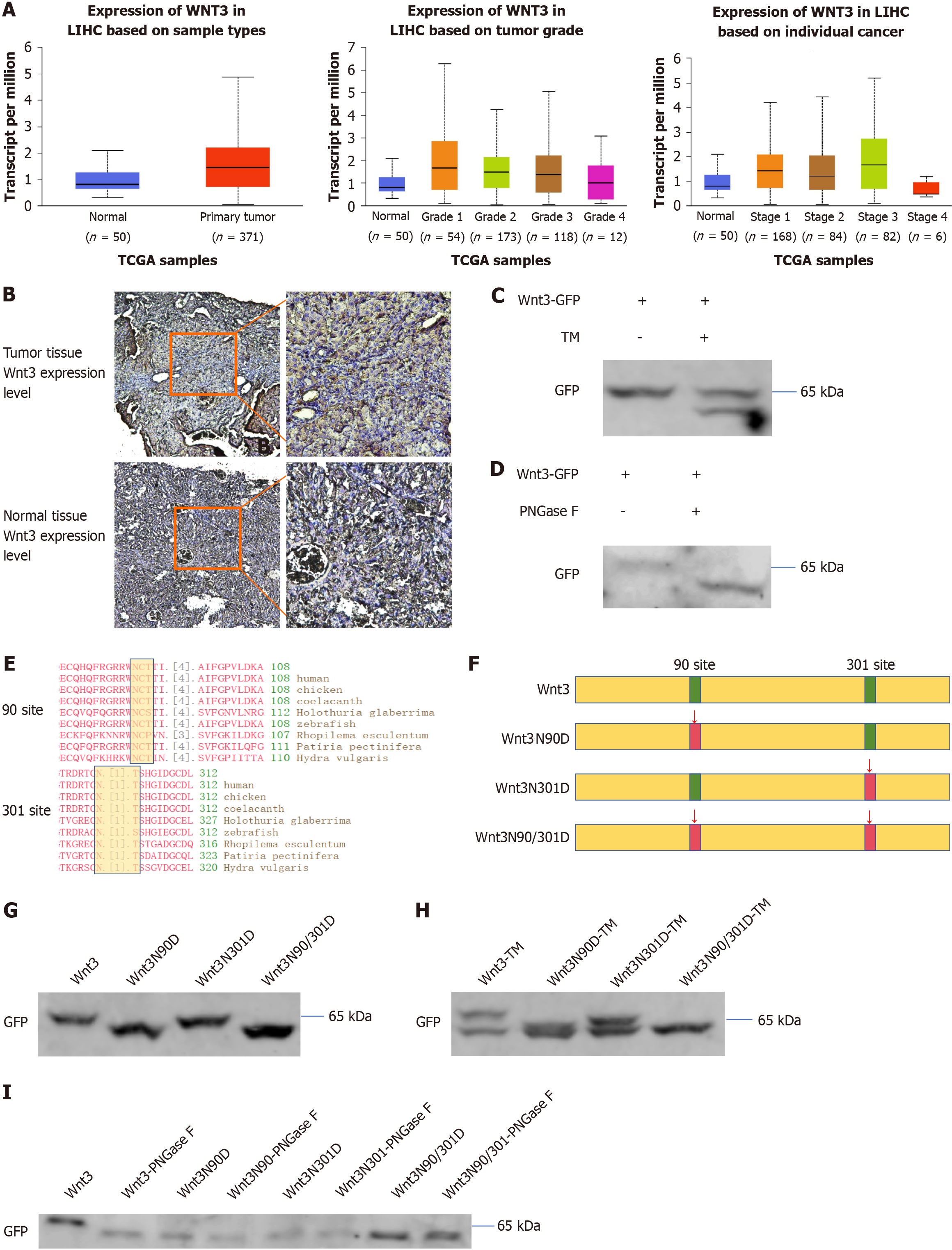
Figure 1 Wnt3 is upregulated in hepatocellular carcinoma and modified by N-glycosylation at residues 90 and 301.
A: According to the TCGA database query, the expression level of Wnt3 increased during the early stage of hepatocellular carcinoma; B: DEN and TAA were used to induce primary liver cancer in C3H mice, and normal mouse liver tissue was used as a control. The expression of Wnt3 in the two tissues was detected by immunohistochemical staining; C and H: Wnt3-GFP was transfected into PLC/PRF/5 cells for 36 h, which were then treated with TM for 24 h at a working concentration of 2.5 μg/mL; D and I: PLC/PRF/5 cells were transfected with Wnt3-GFP for 48 h, after which the cells were collected. Wnt3 was purified with a GFP tag, and the purified protein was treated according to the instructions of the PNGase F deglycosylation kit; E: Query of conserved areas of Wnt3 identified through the National Center for Biotechnology Information database; F: Schematic diagram showing the construction of site-directed Wnt3 mutants; G: Forty-eight hours after transfection, the cells were collected and subjected to Western blotting. LIHC: Liver hepatocellular carcinoma; GFP: Green fluorescent protein.
- Citation: Zhang XZ, Mo XC, Wang ZT, Sun R, Sun DQ. N-glycosylation of Wnt3 regulates the progression of hepatocellular carcinoma by affecting Wnt/β-catenin signal pathway. World J Gastrointest Oncol 2024; 16(6): 2769-2780
- URL: https://www.wjgnet.com/1948-5204/full/v16/i6/2769.htm
- DOI: https://dx.doi.org/10.4251/wjgo.v16.i6.2769


