Copyright
©The Author(s) 2024.
World J Gastrointest Oncol. May 15, 2024; 16(5): 2241-2252
Published online May 15, 2024. doi: 10.4251/wjgo.v16.i5.2241
Published online May 15, 2024. doi: 10.4251/wjgo.v16.i5.2241
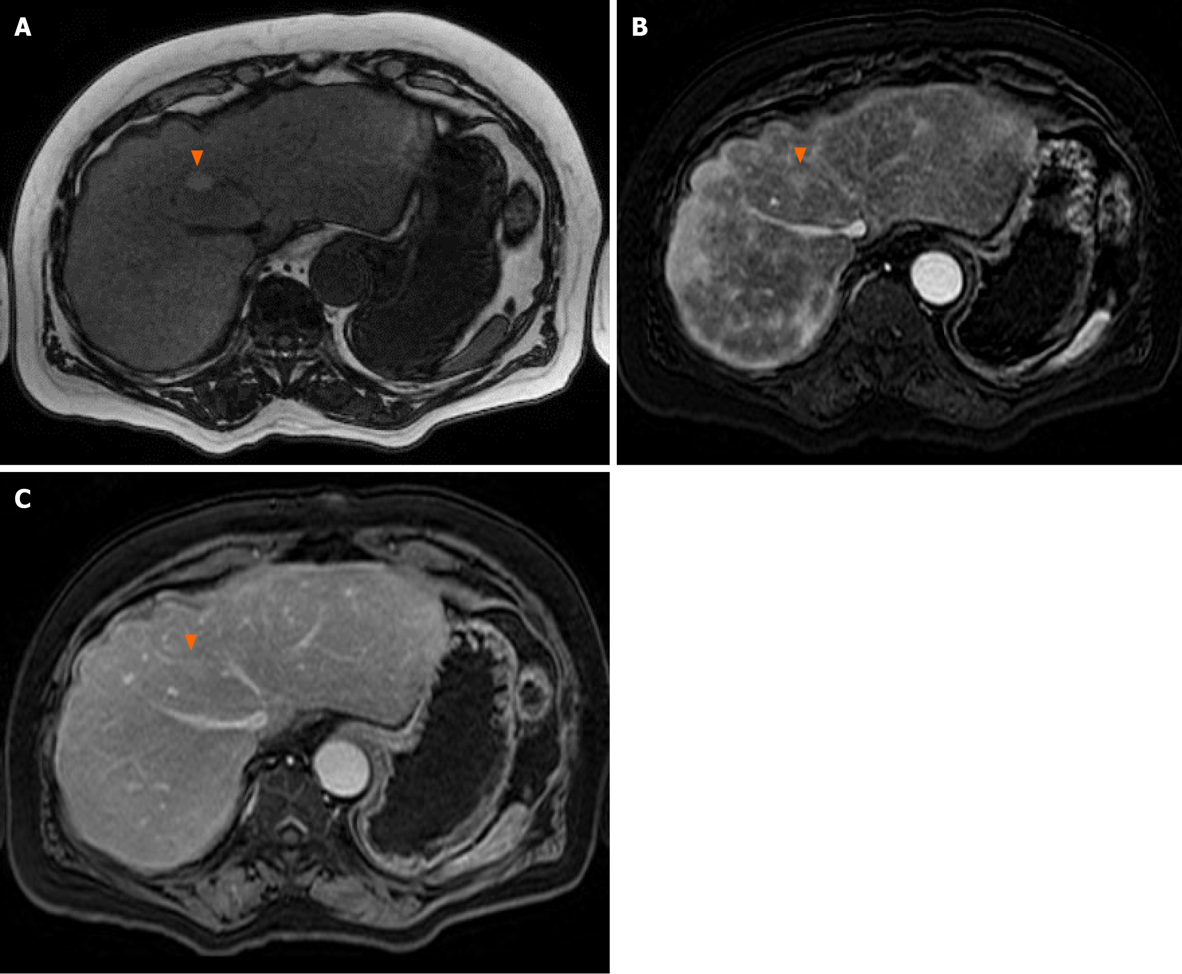
Figure 3 Magnetic resonance imaging of the liver.
Magnetic resonance imaging abdomen demonstrated a 1.2 cm lesion in segment VIII with late arterial enhancement, fatty sparing, and intrinsic T1 hyperintensity. A: T1-weighted, pre-contrast; B: T1-weighted immediate post-contrast; C: T1-weighted, 5 min post-contrast.
- Citation: Wu WK, Patel K, Padmanabhan C, Idrees K. Hepatocellular carcinoma presenting as an extrahepatic mass: A case report and review of literature. World J Gastrointest Oncol 2024; 16(5): 2241-2252
- URL: https://www.wjgnet.com/1948-5204/full/v16/i5/2241.htm
- DOI: https://dx.doi.org/10.4251/wjgo.v16.i5.2241


