Copyright
©The Author(s) 2024.
World J Gastrointest Oncol. May 15, 2024; 16(5): 2241-2252
Published online May 15, 2024. doi: 10.4251/wjgo.v16.i5.2241
Published online May 15, 2024. doi: 10.4251/wjgo.v16.i5.2241
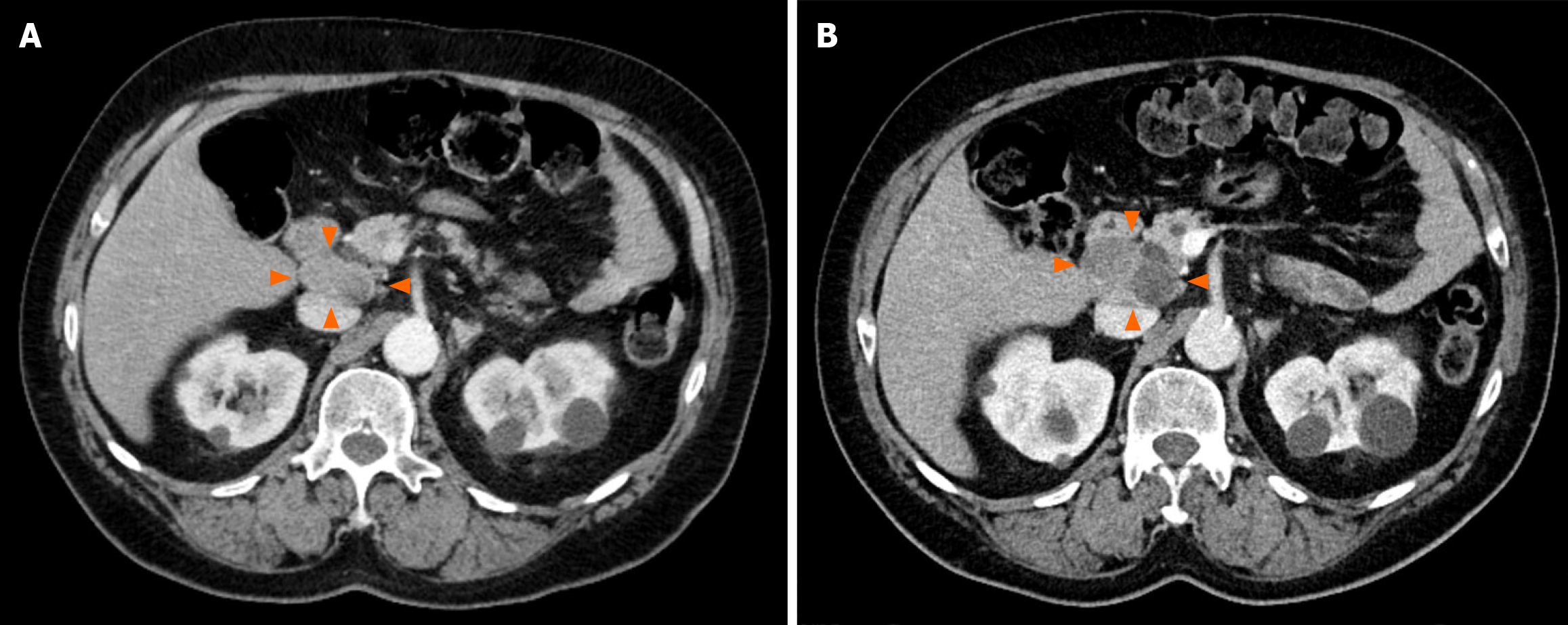
Figure 1 Lesion growth on computed tomography.
Serial intravenous-contrasted Computed tomography scans of the abdomen and pelvis demonstrated increase in size of the patient’s portocaval mass (arrows). A: 3.6 cm × 2 cm, 8 months prior to presentation; B: 5.2 cm × 3.2 cm, time of presentation.
- Citation: Wu WK, Patel K, Padmanabhan C, Idrees K. Hepatocellular carcinoma presenting as an extrahepatic mass: A case report and review of literature. World J Gastrointest Oncol 2024; 16(5): 2241-2252
- URL: https://www.wjgnet.com/1948-5204/full/v16/i5/2241.htm
- DOI: https://dx.doi.org/10.4251/wjgo.v16.i5.2241


