Copyright
©The Author(s) 2024.
World J Gastrointest Oncol. May 15, 2024; 16(5): 2219-2224
Published online May 15, 2024. doi: 10.4251/wjgo.v16.i5.2219
Published online May 15, 2024. doi: 10.4251/wjgo.v16.i5.2219
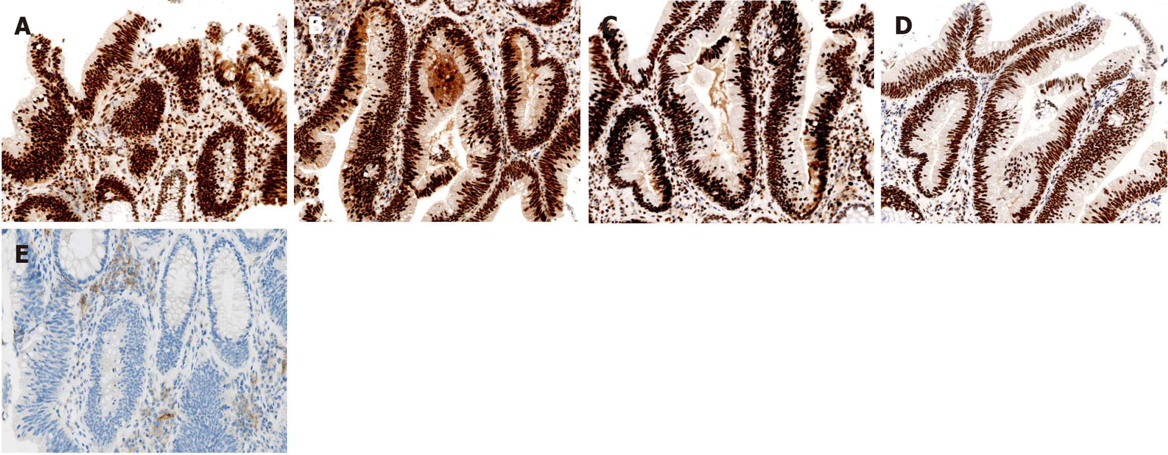
Figure 2 Histopathological analysis and immunohistochemical staining of the biopsy specimen.
A: MLH1 is positive in tumor cells; B: MSH2 is positive in tumor cells; C: MSH6 is positive in tumor cells; D: PMS2 is positive in tumor cells; E: High expression of programmed cell death-ligand 1 in the specimen.
- Citation: Zhong WT, Lv Y, Wang QY, An R, Chen G, Du JF. Chemoradiotherapy plus tislelizumab for mismatch repair proficient rectal cancer with supraclavicular lymph node metastasis: A case report. World J Gastrointest Oncol 2024; 16(5): 2219-2224
- URL: https://www.wjgnet.com/1948-5204/full/v16/i5/2219.htm
- DOI: https://dx.doi.org/10.4251/wjgo.v16.i5.2219


