Copyright
©The Author(s) 2024.
World J Gastrointest Oncol. May 15, 2024; 16(5): 2219-2224
Published online May 15, 2024. doi: 10.4251/wjgo.v16.i5.2219
Published online May 15, 2024. doi: 10.4251/wjgo.v16.i5.2219
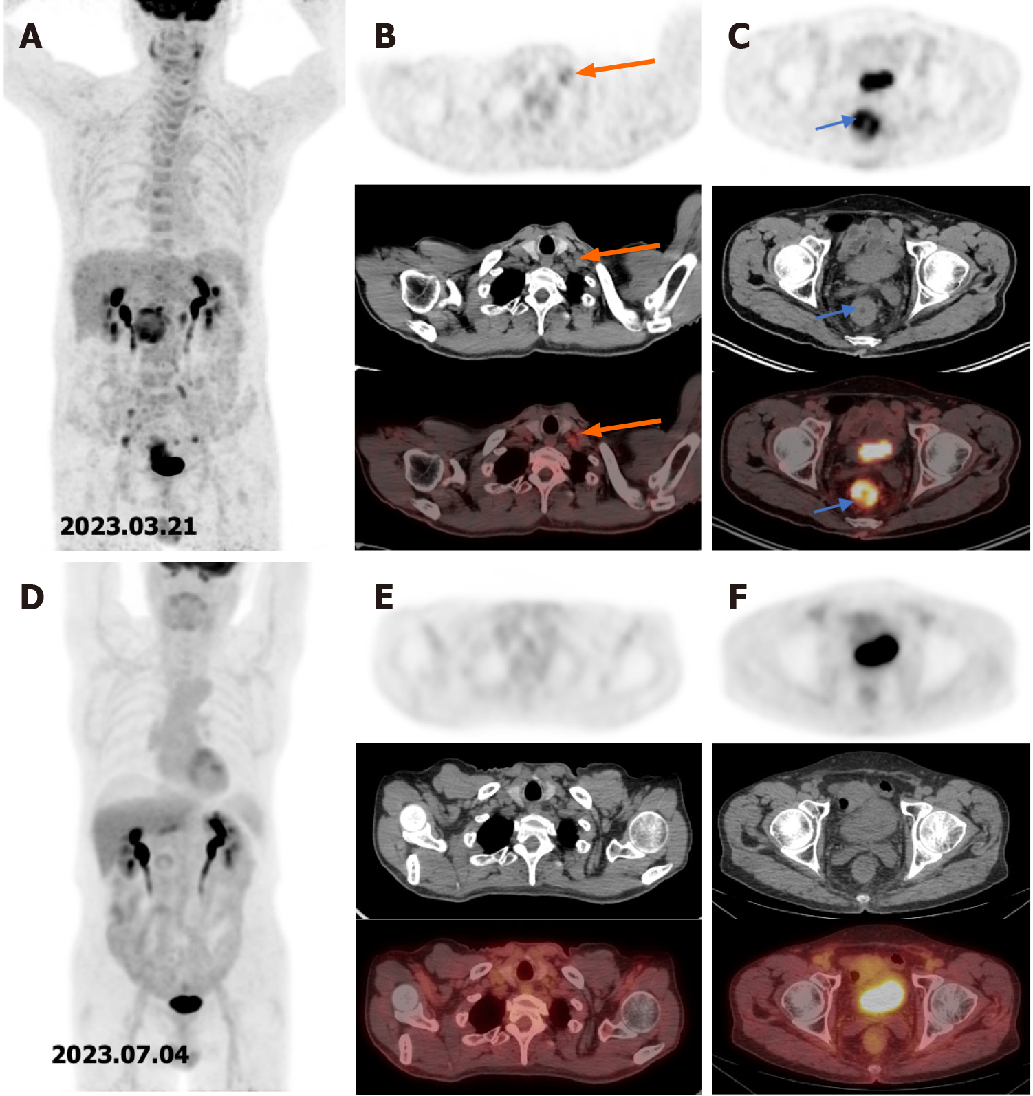
Figure 1 18F-FDG PET/CT image before and after treatment.
A: 18F-FDG PET/CT image before treatment. The number is time of examination; B: Metastatic lymph node in the clavicle before treatment. The orange arrow indicates the lymph node; C: The rectal lesion before treatment. The lesion is indicated by the blue arrows; D: 18F-FDG PET/CT image after treatment. The number is time of examination; E: Disappearance of the metastatic lymph node after treatment; F: The rectal lesion with reduced uptake after treatment.
- Citation: Zhong WT, Lv Y, Wang QY, An R, Chen G, Du JF. Chemoradiotherapy plus tislelizumab for mismatch repair proficient rectal cancer with supraclavicular lymph node metastasis: A case report. World J Gastrointest Oncol 2024; 16(5): 2219-2224
- URL: https://www.wjgnet.com/1948-5204/full/v16/i5/2219.htm
- DOI: https://dx.doi.org/10.4251/wjgo.v16.i5.2219


