Copyright
©The Author(s) 2024.
World J Gastrointest Oncol. May 15, 2024; 16(5): 1861-1868
Published online May 15, 2024. doi: 10.4251/wjgo.v16.i5.1861
Published online May 15, 2024. doi: 10.4251/wjgo.v16.i5.1861
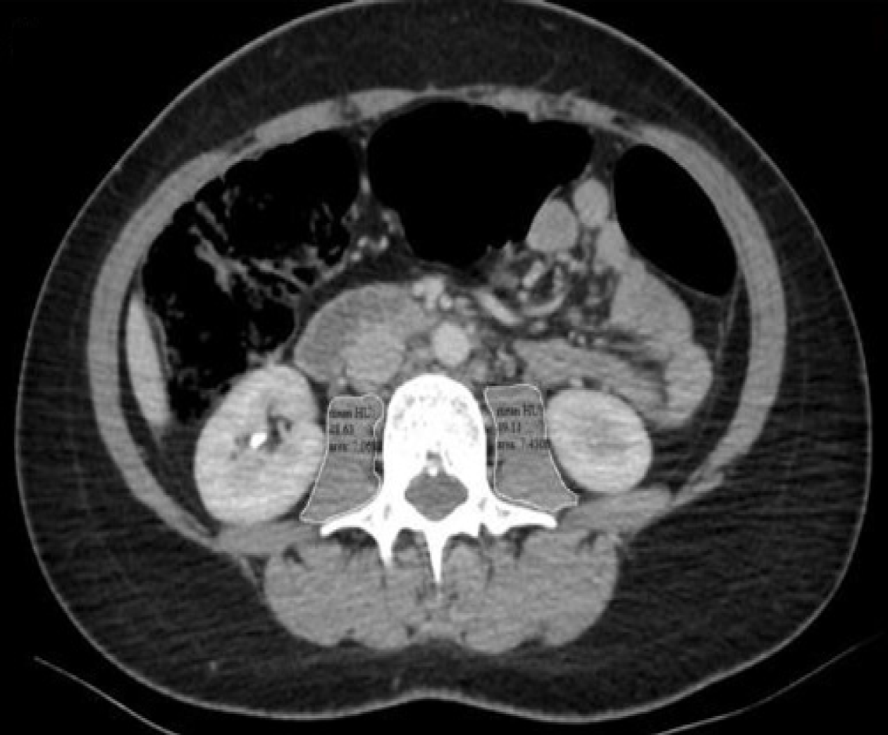
Figure 1 Abdominal axial computed tomography images.
At the level of third lumbar vertebra, outer boundaries of psoas muscles were drawn manually (white lines) and psoas muscle cross sectional area (cm2) and attenuation (Hounsfield unit) were measured on both sides.
- Citation: Dogan O, Sahinli H, Duzkopru Y, Akdag T, Kocanoglu A. Is sarcopenia effective on survival in patients with metastatic gastric cancer? World J Gastrointest Oncol 2024; 16(5): 1861-1868
- URL: https://www.wjgnet.com/1948-5204/full/v16/i5/1861.htm
- DOI: https://dx.doi.org/10.4251/wjgo.v16.i5.1861


