Copyright
©The Author(s) 2024.
World J Gastrointest Oncol. May 15, 2024; 16(5): 1821-1832
Published online May 15, 2024. doi: 10.4251/wjgo.v16.i5.1821
Published online May 15, 2024. doi: 10.4251/wjgo.v16.i5.1821
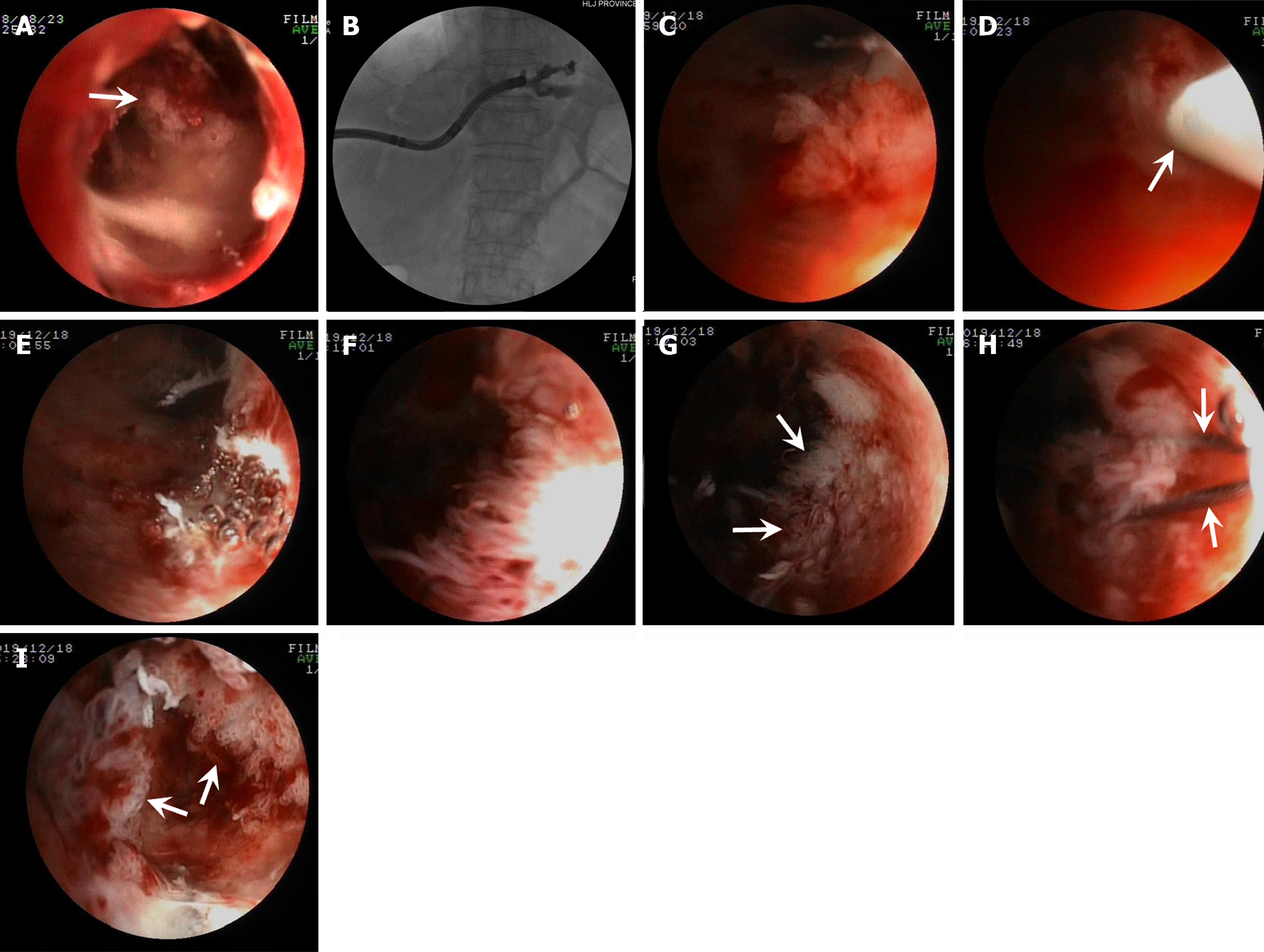
Figure 5 Percutaneous transhepatic cholangioscopy-assisted biliary polypectomy for mucin-hypersecreting cast-like type intraductal papillary neoplasm in the intrahepatic bile duct, with technical success in patient five.
A: Percutaneous transhepatic cholangioscopy (PTCS) showing the biliary tumors at the proximal end of the stricture; B: Cholangiography showing the location of the stricture in the left intrahepatic bile duct; C: PTCS showed a frond-like tumor; D: The snared tumor (the arrow points to the snare sheath); E: The successfully resected tumor with PTCS-assisted biliary polypectomy (PTCS-BP); F: PTCS showing multiple villous growing tumors; G: PTCS showing some residual tumors (arrows) after PTCS-BP; H: Subsequent PTCS-BP performed for the residual villous growing tumors (arrows point to the snare steel wire); I: PTCS showing unresected residual tumors (arrows) in the small ductal lumen.
- Citation: Ren X, Qu YP, Zhu CL, Xu XH, Jiang H, Lu YX, Xue HP. Percutaneous transhepatic cholangioscopy-assisted biliary polypectomy for local palliative treatment of intraductal papillary neoplasm of the bile duct. World J Gastrointest Oncol 2024; 16(5): 1821-1832
- URL: https://www.wjgnet.com/1948-5204/full/v16/i5/1821.htm
- DOI: https://dx.doi.org/10.4251/wjgo.v16.i5.1821


