Copyright
©The Author(s) 2024.
World J Gastrointest Oncol. May 15, 2024; 16(5): 1821-1832
Published online May 15, 2024. doi: 10.4251/wjgo.v16.i5.1821
Published online May 15, 2024. doi: 10.4251/wjgo.v16.i5.1821
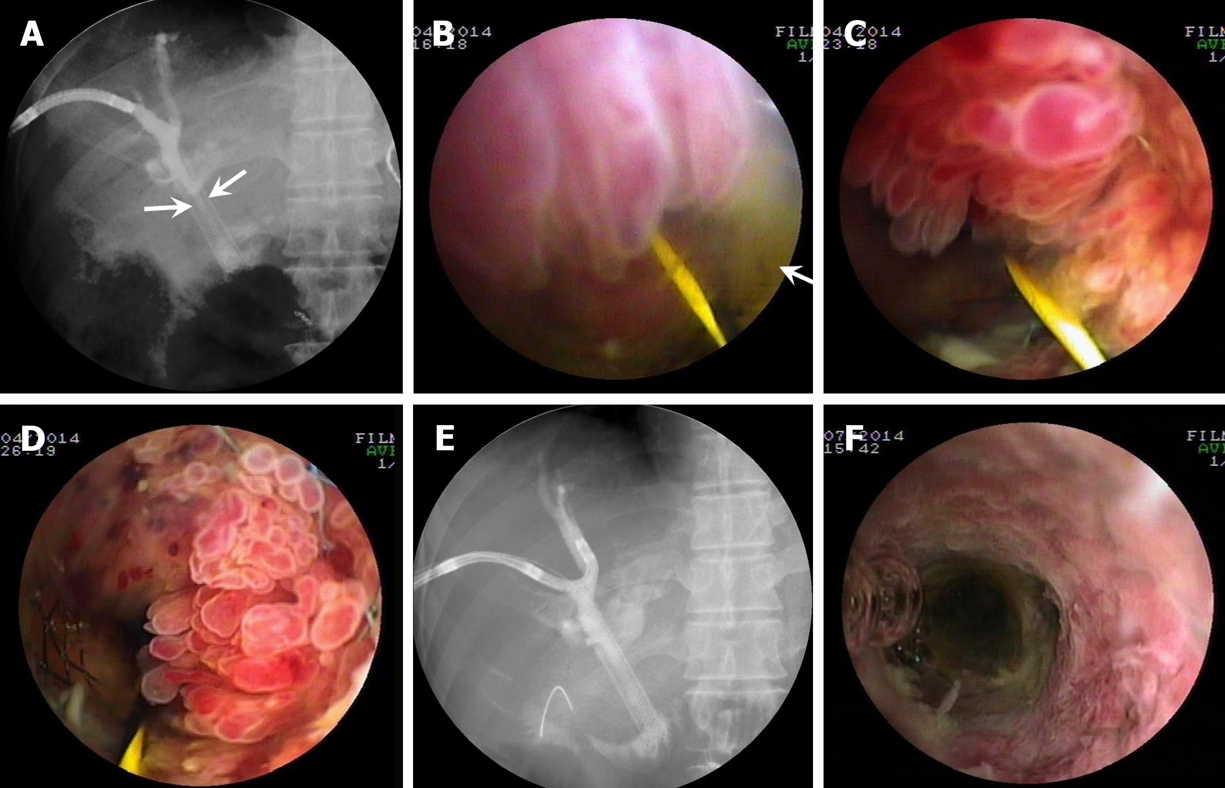
Figure 4 Percutaneous transhepatic cholangioscopy-assisted biliary polypectomy for postoperative recurrent mucin-hypersecreting cast-like type intraductal papillary neoplasm of the bile duct, with therapeutic success in patient four who received transanastomotic metal stenting for intraductal papillary neoplasm of the bile duct one month ago.
A: Cholangiography showing stenosis of the metal stent lumen (arrows); B-D: Percutaneous transhepatic cholangioscopy (PTCS) showed thick mucus (arrow) and multiple villous, frond-like, and fish-egg like protrusions in the stent lumen, respectively; E: Cholangiography showing the improvement of the stricture with a patent metal stent in the lumen; F: Repeated PTCS showing no obvious elevated tumor regrowth in the metal stent 3 months after PTCS-assisted biliary polypectomy.
- Citation: Ren X, Qu YP, Zhu CL, Xu XH, Jiang H, Lu YX, Xue HP. Percutaneous transhepatic cholangioscopy-assisted biliary polypectomy for local palliative treatment of intraductal papillary neoplasm of the bile duct. World J Gastrointest Oncol 2024; 16(5): 1821-1832
- URL: https://www.wjgnet.com/1948-5204/full/v16/i5/1821.htm
- DOI: https://dx.doi.org/10.4251/wjgo.v16.i5.1821


