Copyright
©The Author(s) 2024.
World J Gastrointest Oncol. May 15, 2024; 16(5): 1821-1832
Published online May 15, 2024. doi: 10.4251/wjgo.v16.i5.1821
Published online May 15, 2024. doi: 10.4251/wjgo.v16.i5.1821
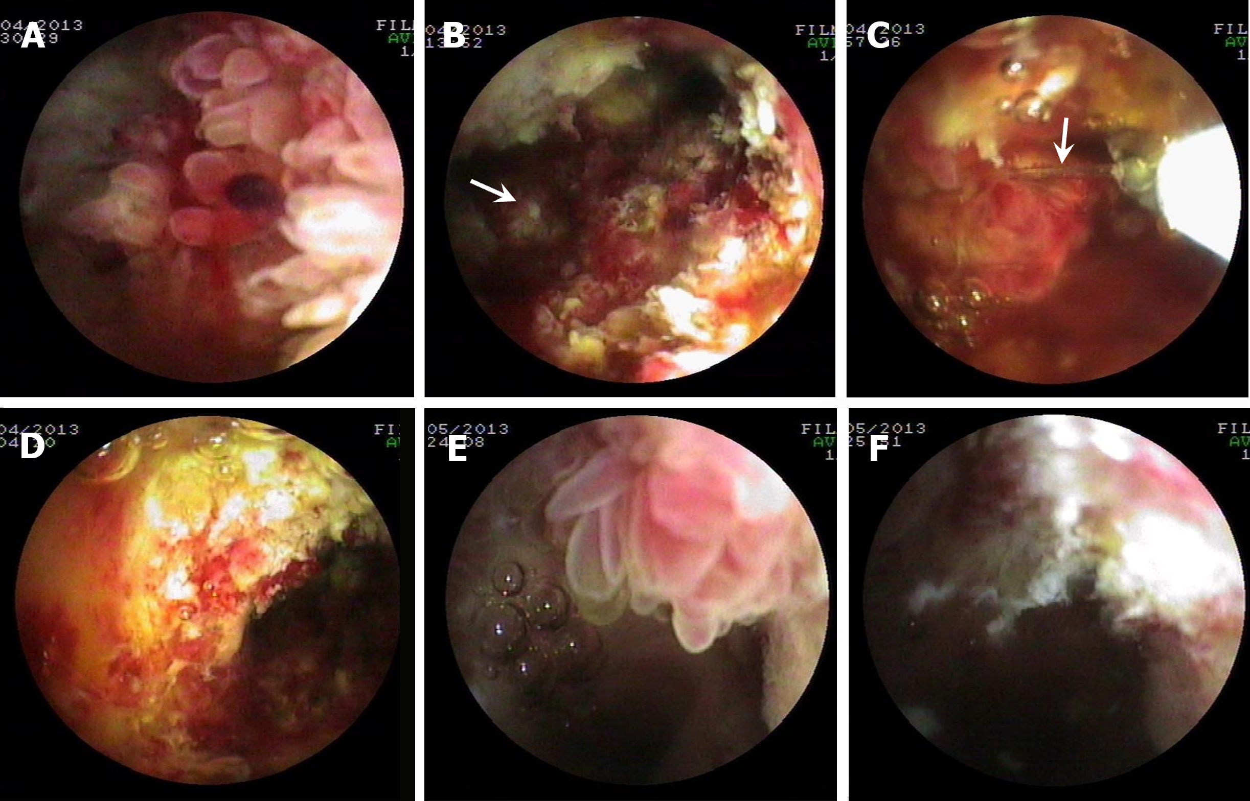
Figure 2 Percutaneous transhepatic cholangioscopy-assisted biliary polypectomy for recurrent mucin-hypersecreting cast-like type intraductal papillary neoplasm of the bile duct with therapeutic success in patient two who received photodynamic therapy for postoperative recurrent intraductal papillary neoplasm of the bile duct 11 months prior to Percutaneous transhepatic cholangioscopy-assisted biliary polypectomy.
A and B: Percutaneous transhepatic cholangioscopy (PTCS) showing the multiple papillary tumors and the locations (the arrow in Figure B refers to a resected piece of the tumor tissue) where the tumors were resected with PTCS-assisted biliary polypectomy (PTCS-BP), respectively; C and D: PTCS showing a polypoid tumor snared (the arrow in Figure C points to the snare steel wire) and the location where the tumor was resected with PTCS-BP, respectively; E and F: PTCS showing a frond-like projection and the locations where the tumors were resected with PTCS-BP, respectively; A, C, and E: PTCS showing the multiple papillary, a polypoid and frond-like tumors, respectively; B, D, and F: PTCS showing the locations where the tumor was successfully resected with PTCS-BP.
- Citation: Ren X, Qu YP, Zhu CL, Xu XH, Jiang H, Lu YX, Xue HP. Percutaneous transhepatic cholangioscopy-assisted biliary polypectomy for local palliative treatment of intraductal papillary neoplasm of the bile duct. World J Gastrointest Oncol 2024; 16(5): 1821-1832
- URL: https://www.wjgnet.com/1948-5204/full/v16/i5/1821.htm
- DOI: https://dx.doi.org/10.4251/wjgo.v16.i5.1821


