Copyright
©The Author(s) 2024.
World J Gastrointest Oncol. May 15, 2024; 16(5): 1821-1832
Published online May 15, 2024. doi: 10.4251/wjgo.v16.i5.1821
Published online May 15, 2024. doi: 10.4251/wjgo.v16.i5.1821
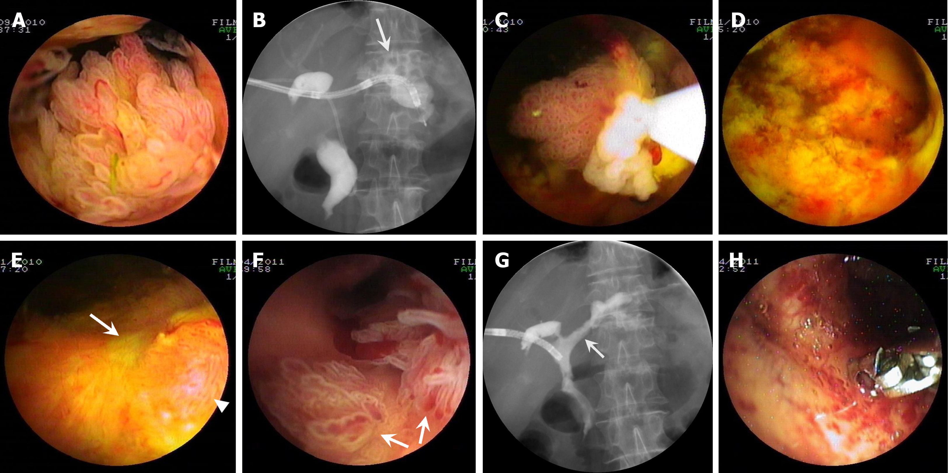
Figure 1 Percutaneous transhepatic cholangioscopy-assisted biliary polypectomy for mucin-hypersecreting cast-like type intraductal papillary neoplasm of the bile duct with therapeutic success in patient one.
A: Percutaneous transhepatic cholangioscopy (PTCS) showing frond-like tumors with vascular images in the hilar bile duct; B: Cholangiography showing multiple 1-cm sized stones in the left intrahepatic duct (arrow); C: PTCS showing a frond-like tumor that was being resected with the snare at the lower right in the visual field under PTCS; D: The cholangioscopic view of the location where the tumor was successfully resected with PTCS assisted biliary polypectomy (PTCS-BP); E: A scar (arrow) forming with a local minimal proliferated tumor (arrowhead) 2 wk after the last PTCS-BP; F: PTCS showing newly growing leaf-like tumors (arrows); G: Cholangiography showing a reduced biliary tract caliber, with a stenotic stiff and rigid ductal wall suspected of the existence of an invasive lesion in the left hepatic duct (arrow); H: Biopsies being taken from the site suspected of the existence of an invasive lesion 5 months after PTCS-BP.
- Citation: Ren X, Qu YP, Zhu CL, Xu XH, Jiang H, Lu YX, Xue HP. Percutaneous transhepatic cholangioscopy-assisted biliary polypectomy for local palliative treatment of intraductal papillary neoplasm of the bile duct. World J Gastrointest Oncol 2024; 16(5): 1821-1832
- URL: https://www.wjgnet.com/1948-5204/full/v16/i5/1821.htm
- DOI: https://dx.doi.org/10.4251/wjgo.v16.i5.1821


