Copyright
©The Author(s) 2024.
World J Gastrointest Oncol. May 15, 2024; 16(5): 1773-1786
Published online May 15, 2024. doi: 10.4251/wjgo.v16.i5.1773
Published online May 15, 2024. doi: 10.4251/wjgo.v16.i5.1773
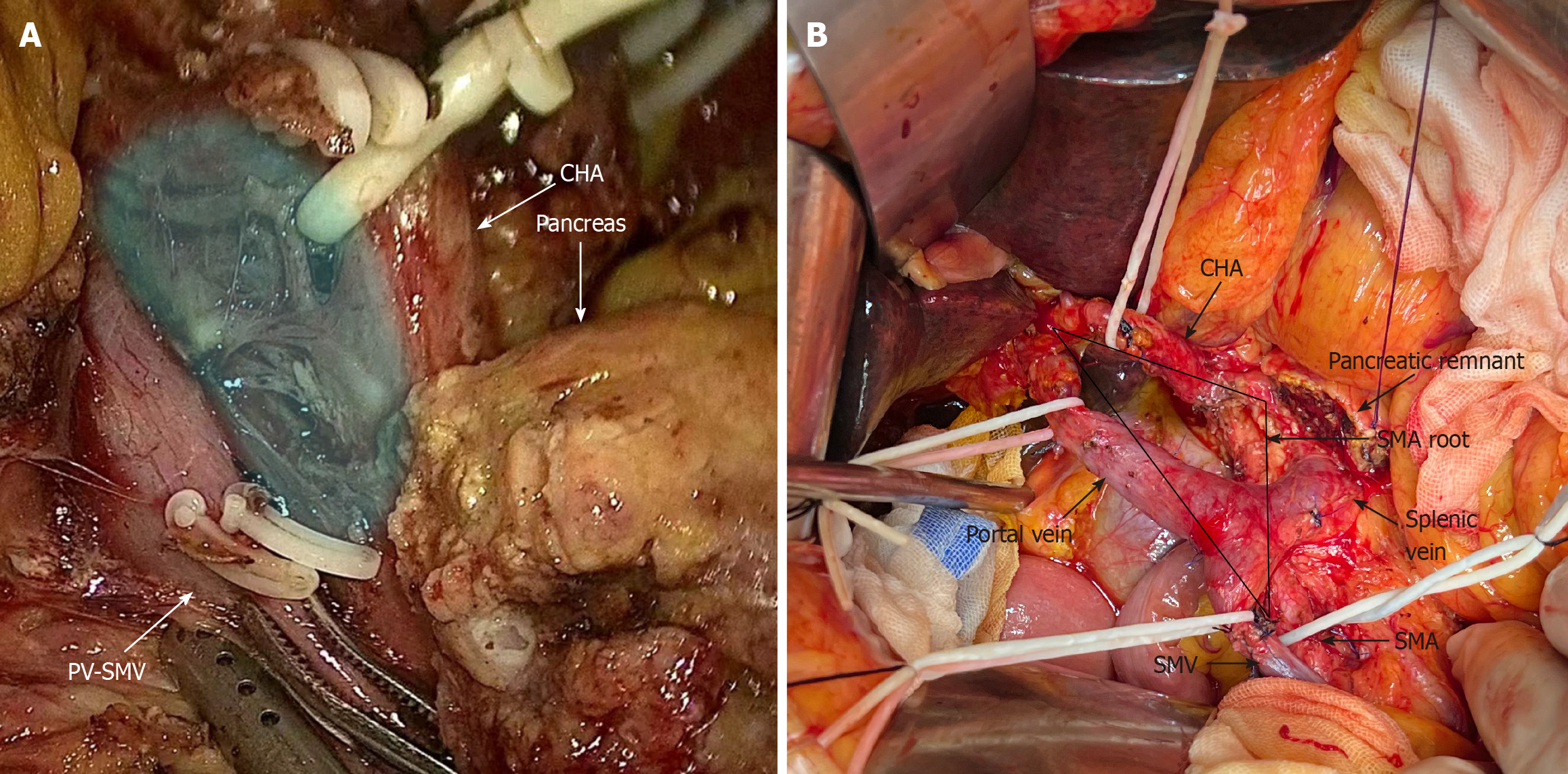
Figure 2 Intraoperative image of the Heidelberg triangle dissection.
A: Intraoperative image before Heidelberg triangle dissection. At this stage, the superior mesenteric artery (SMA) is located on the dorsal side of the pancreatic remnant, and the portal vein (PV)-superior mesenteric vein (SMV), common hepatic artery (CHA), and pancreas are visible. The blue shading illustrates the boundaries of the Heidelberg triangle, encompassing vascular, lymphatic, and neural fiber tissues within the triangle; B: Intraoperative image after Heidelberg triangle dissection. The black arrowhead points to vessels and tissues around the Heidelberg triangle. The black triangle shows the range of Heidelberg triangle dissection. After thorough clearance, the precise boundaries of the triangle and its three bordering vessels, namely, the PV-SMV, CHA, and SMA, are clearly visible. SMV: Superior mesenteric vein; SMA: Superior mesenteric artery; CHA: Common hepatic artery; PV: Portal vein.
- Citation: Chen JH, Zhu LY, Cai ZW, Hu X, Ahmed AA, Ge JQ, Tang XY, Li CJ, Pu YL, Jiang CY. TRIANGLE operation, combined with adequate adjuvant chemotherapy, can improve the prognosis of pancreatic head cancer: A retrospective study. World J Gastrointest Oncol 2024; 16(5): 1773-1786
- URL: https://www.wjgnet.com/1948-5204/full/v16/i5/1773.htm
- DOI: https://dx.doi.org/10.4251/wjgo.v16.i5.1773


