Copyright
©The Author(s) 2024.
World J Gastrointest Oncol. May 15, 2024; 16(5): 1773-1786
Published online May 15, 2024. doi: 10.4251/wjgo.v16.i5.1773
Published online May 15, 2024. doi: 10.4251/wjgo.v16.i5.1773
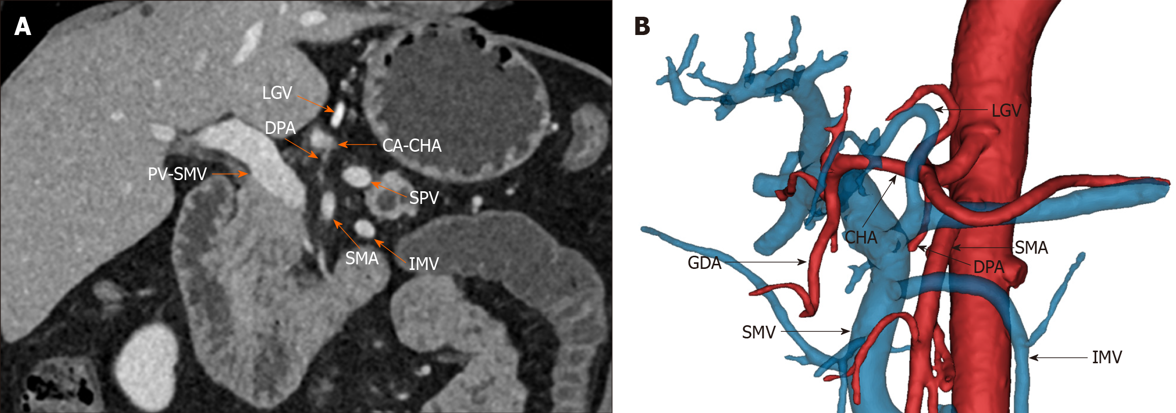
Figure 1 The preoperative imaging of Heidelberg triangle.
A: Preoperative contrast-enhanced computed tomography image of the abdomen. The orange arrowhead points to vessels around the Heidelberg triangle. The portal vein-superior mesenteric vein, celiac axis-common hepatic artery (CHA), and superior mesenteric artery represent the three boundary vessels of the Heidelberg triangle. The dorsal pancreatic artery (DPA) in the figure originates from the CHA and passes through the triangle, reflecting one of the numerous pathways of DPA. The left gastric vein (LGV), splenic vein (SPV), and inferior mesenteric vein (IMV) typically pass anteriorly to the triangle; B: Frontal view of the Heidelberg triangle generated by three-dimensional reconstruction. The black arrowhead points to vessels around the Heidelberg triangle. The image reveals the DPA coursing within the triangle and the LGV, SPV, and IMV traversing anteriorly to the triangle. GDA: Gastroduodenal artery; PV: Portal vein; SMV: Superior mesenteric vein; CA: Celiac axis; CHA: Common hepatic artery; SMA: Superior mesenteric artery; DPA: Dorsal pancreatic artery; LGV: Left gastric vein; SPV: Splenic vein; IMV: Inferior mesenteric vein.
- Citation: Chen JH, Zhu LY, Cai ZW, Hu X, Ahmed AA, Ge JQ, Tang XY, Li CJ, Pu YL, Jiang CY. TRIANGLE operation, combined with adequate adjuvant chemotherapy, can improve the prognosis of pancreatic head cancer: A retrospective study. World J Gastrointest Oncol 2024; 16(5): 1773-1786
- URL: https://www.wjgnet.com/1948-5204/full/v16/i5/1773.htm
- DOI: https://dx.doi.org/10.4251/wjgo.v16.i5.1773


